Thomas Schultz Gemma Nedjati-Gilani Archana Venkataraman - Computational Diffusion MRI and Brain Connectivity
Here you can read online Thomas Schultz Gemma Nedjati-Gilani Archana Venkataraman - Computational Diffusion MRI and Brain Connectivity full text of the book (entire story) in english for free. Download pdf and epub, get meaning, cover and reviews about this ebook. City: Cham, year: 2016, publisher: Springer International Publishing, genre: Home and family. Description of the work, (preface) as well as reviews are available. Best literature library LitArk.com created for fans of good reading and offers a wide selection of genres:
Romance novel
Science fiction
Adventure
Detective
Science
History
Home and family
Prose
Art
Politics
Computer
Non-fiction
Religion
Business
Children
Humor
Choose a favorite category and find really read worthwhile books. Enjoy immersion in the world of imagination, feel the emotions of the characters or learn something new for yourself, make an fascinating discovery.

- Book:Computational Diffusion MRI and Brain Connectivity
- Author:
- Publisher:Springer International Publishing
- Genre:
- Year:2016
- City:Cham
- Rating:3 / 5
- Favourites:Add to favourites
- Your mark:
Computational Diffusion MRI and Brain Connectivity: summary, description and annotation
We offer to read an annotation, description, summary or preface (depends on what the author of the book "Computational Diffusion MRI and Brain Connectivity" wrote himself). If you haven't found the necessary information about the book — write in the comments, we will try to find it.
Thomas Schultz Gemma Nedjati-Gilani Archana Venkataraman: author's other books
Who wrote Computational Diffusion MRI and Brain Connectivity? Find out the surname, the name of the author of the book and a list of all author's works by series.


Computational Diffusion MRI and Brain Connectivity — read online for free the complete book (whole text) full work
Below is the text of the book, divided by pages. System saving the place of the last page read, allows you to conveniently read the book "Computational Diffusion MRI and Brain Connectivity" online for free, without having to search again every time where you left off. Put a bookmark, and you can go to the page where you finished reading at any time.
Font size:
Interval:
Bookmark:
Acquisition of Diffusion MRI
 penalty on SNR (where R is the acceleration factor) found in conventional parallel imaging acceleration. Thus, in this work, we use the multi-slice acquisition protocol detailed in [], which uses both the inplane (acceleration factor of 2) and slice accelerations to simultaneously acquire data from multiple slices. In this case, the repetition time TR is reduced proportionately to the number of multi-slice acquisitions R .
 is the gradient direction,
  and w i forms the volume fraction of each component, D 1 and D 2 are cylindrical tensors and D iso is an isotropic tensor with fixed diffusivity of 0.003mm2s as given in [).
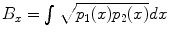 , where p 1( x ), p 2( x ) are the pdfs to be compared. To compute the distance between two fiber bundles, we simply take an equally-weighted combination in each co-ordinate:
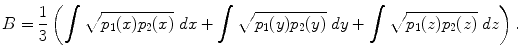
 . Thus, values of B are bounded between 0 and 1. Further, B will be 1 for a perfect match between two fiber bundles and 0 for no overlap at all.
Font size:
Interval:
Bookmark:
Similar books «Computational Diffusion MRI and Brain Connectivity»
Look at similar books to Computational Diffusion MRI and Brain Connectivity. We have selected literature similar in name and meaning in the hope of providing readers with more options to find new, interesting, not yet read works.
Discussion, reviews of the book Computational Diffusion MRI and Brain Connectivity and just readers' own opinions. Leave your comments, write what you think about the work, its meaning or the main characters. Specify what exactly you liked and what you didn't like, and why you think so.