• Complain

Michael Kyba - Skeletal Muscle Regeneration in the Mouse: Methods and Protocols

Here you can read online Michael Kyba - Skeletal Muscle Regeneration in the Mouse: Methods and Protocols full text of the book (entire story) in english for free. Download pdf and epub, get meaning, cover and reviews about this ebook. year: 2016, publisher: Springer, genre: Home and family. Description of the work, (preface) as well as reviews are available. Best literature library LitArk.com created for fans of good reading and offers a wide selection of genres:

Romance novel Science fiction Adventure Detective Science History Home and family Prose Art Politics Computer Non-fiction Religion Business Children Humor

Choose a favorite category and find really read worthwhile books. Enjoy immersion in the world of imagination, feel the emotions of the characters or learn something new for yourself, make an fascinating discovery.

Michael Kyba Skeletal Muscle Regeneration in the Mouse: Methods and Protocols
  • Book:
    Skeletal Muscle Regeneration in the Mouse: Methods and Protocols
  • Author:
  • Publisher:
    Springer
  • Genre:
  • Year:
    2016
  • Rating:
    3 / 5
  • Favourites:
    Add to favourites
  • Your mark:
    • 60
    • 1
    • 2
    • 3
    • 4
    • 5

Skeletal Muscle Regeneration in the Mouse: Methods and Protocols: summary, description and annotation

We offer to read an annotation, description, summary or preface (depends on what the author of the book "Skeletal Muscle Regeneration in the Mouse: Methods and Protocols" wrote himself). If you haven't found the necessary information about the book — write in the comments, we will try to find it.

This volume focuses on the cell biology and physiology of skeletal muscle regeneration. This Book is a collection of classic and cutting edge protocols optimized for mice, but in most cases adaptable to rat or other mammalian models, that will allow an investigator to develop and implement a research study on skeletal muscle regeneration. Chapters address the three major areas of study: provoking regeneration by inducing damage to muscle, analyzing the progenitor cells of skeletal muscle, and quantifying overall muscle function. Subjects discussed include: inducing skeletal muscle injury by eccentric contraction; volumetric muscle loss; single myofiber isolation and culture; satellite cell transplantation; muscle clearing for whole mount immunostaining; luciferase tracking of muscle stem cells; mitochondrial and mitophagy flux analysis; in vivo assessment of muscle contractility; force measurements on single isolated myofibers; and analysis of aerobic respiration in intact skeletal muscle tissue by microplate respirometry. Written in the highly successful Methods in Molecular Biology series format, chapters include introductions to each respective topic, lists of the necessary materials and reagents, step-by-step, readily reproducible laboratory protocols, and tips on troubleshooting and avoiding known pitfalls.
Cutting edge and practical, Skeletal Muscle Regeneration in the Mouse: Methods and Protocols is an essential laboratory reference for research in skeletal muscle growth, damage, repair, degeneration, and regenerative therapy in the mouse model system.

Michael Kyba: author's other books


Who wrote Skeletal Muscle Regeneration in the Mouse: Methods and Protocols? Find out the surname, the name of the author of the book and a list of all author's works by series.

Skeletal Muscle Regeneration in the Mouse: Methods and Protocols — read online for free the complete book (whole text) full work

Below is the text of the book, divided by pages. System saving the place of the last page read, allows you to conveniently read the book "Skeletal Muscle Regeneration in the Mouse: Methods and Protocols" online for free, without having to search again every time where you left off. Put a bookmark, and you can go to the page where you finished reading at any time.

Light

Font size:

Reset

Interval:

Bookmark:

Make
Part I
Injury Models
Springer Science+Business Media New York 2016
Michael Kyba (ed.) Skeletal Muscle Regeneration in the Mouse Methods in Molecular Biology 1460 10.1007/978-1-4939-3810-0_1
1. Eccentric Contraction-Induced Muscle Injury: Reproducible, Quantitative, Physiological Models to Impair Skeletal Muscles Capacity to Generate Force
Jarrod A. Call 1, 2 and Dawn A. Lowe 3
(1)
Department of Kinesiology, University of Georgia, Athens, GA 30602, USA
(2)
Regenerative Bioscience Center, University of Georgia, Athens, GA 30602, USA
(3)
Programs in Physical Therapy and Rehabilitation Science, Department of Physical Medicine and Rehabilitation Medical School, University of Minnesota, Minneapolis, MN 55455, USA
Dawn A. Lowe
Email:
Abstract
In order to investigate the molecular and cellular mechanisms of muscle regeneration an experimental injury model is required. Advantages of eccentric contraction-induced injury are that it is a controllable, reproducible, and physiologically relevant model to cause muscle injury, with injury being defined as a loss of force generating capacity. While eccentric contractions can be incorporated into conscious animal study designs such as downhill treadmill running, electrophysiological approaches to elicit eccentric contractions and examine muscle contractility, for example before and after the injurious eccentric contractions, allows researchers to circumvent common issues in determining muscle function in a conscious animal (e.g., unwillingness to participate). Herein, we describe in vitro and in vivo methods that are reliable, repeatable, and truly maximal because the muscle contractions are evoked in a controlled, quantifiable manner independent of subject motivation. Both methods can be used to initiate eccentric contraction-induced injury and are suitable for monitoring functional muscle regeneration hours to days to weeks post-injury.
Key words
Force drop Lengthening contraction Muscle damage
Introduction
Eccentric contractions are contractions in which the external load or resistance placed on an activated muscle is greater than the force generated by that muscle, and subsequently the muscle is lengthened while it is active. There is an immediate loss of strength following the performance of eccentric contractions that is attributed to disruption of the excitationcontraction coupling process and/or frank damage to force-generating or -transmitting structures within the muscle []. The extent of the strength loss can vary depending on age and disease of the subject. For example, acute strength loss can be as high as 50% in muscles of healthy C57Bl mice, but in excess of 75% in an mdx mouse (model for Duchenne muscular dystrophy).
In vitro and in vivo methods are ideal for executing the eccentric contraction-induced injury model because the severity of muscle injury can be monitored in real time and controlled precisely by altering the number of eccentric contractions performed, the distance the muscle is lengthened, and/or the velocity at which the lengthening occurs. Additionally, these methods provide a level of consistency as far as injury at the fiber level because all motor units are activated which is in sharp contrast to fully conscious experimental models, e.g., downhill treadmill running.
1.1 In Vitro Eccentric Contractions
Live, isolated muscle preparations are utilized to assess contractile capacity of those organs. Small muscles of mice, such as the soleus and extensor digitorum longus (EDL) muscles, are suitable and most commonly used because oxygen diffusion to fibers in the core of muscle is adequate [], a host of contractile tests can be performed. The in vitro section of this chapter will focus on eccentric contractions by isolated muscles and the consequent contraction-induced injury. In the muscular dystrophy field, the term force drop is becoming common. This term is used to describe the outcome of eccentric contraction testing in EDL muscle lacking dystrophin as these muscles are highly susceptible to injury, as measured by the loss of force-generating capacity during and immediately following eccentric contractions. Before detailing methods for an in vitro eccentric contraction-injury protocol, a description of EDL muscle dissection is presented in this chapter because careful dissection is important in order to study contraction-induced injury to the muscle as opposed to dissection-induced injury.
1.2 In Vivo Eccentric Contractions
We have developed a modified in vivo muscle testing apparatus and protocol based on the original in vivo system reported by Ashton-Miller et al. []. Briefly, peak isometric contractility of either the ankle dorsi-flexors (tibialis anterior, extensor digitorum longus, extensor hallicus longus muscles) or plantar-flexors (gastrocnemius, soleus, plantaris muscles) in anesthetized mice is determined using percutaneous electrodes to stimulate specific nerves innervating those muscle groups and specialized equipment to record the contractile output. Then an eccentric contraction-induced injury protocol is performed to cause muscle injury as immediately measurable by decrements in torque. Below we detail our eccentric contraction-induced injury model including internal controls and expected outcomes for healthy C57Bl6 mice and some examples from dystrophic mdx mice. Finally, a real advantage of the relatively noninvasive muscle analysis in vivo is that muscle contractile function can be measured repeatedly which can be ideal for monitoring ongoing muscle regeneration. Accordingly, we also briefly describe the recovery of muscle strength after eccentric contraction-induced injury.
Materials
In Vitro
2.1 Equipment and Surgical Instruments for Delicate Muscle Dissections
  1. Dissecting microscope (e.g., Leica S6D).
  2. Fiber-Lite Dual Gooseneck Light (this is needed if there is not a light ring on dissecting scope).
  3. Halstead Mosquito Forceps (~hemostat), curved, delicate 5 length (e.g., George Tiemann #105-1107).
  4. Dressing and Tissue Forceps, delicate with teeth (e.g., George Tiemann #105-205-1).
  5. Extra fine Graefe Forceps, curved, finely grated tips (Fine Science #11151-10).
  6. Dumont Forceps, pointed tip (e.g., Fine Science #11295-10).
  7. Tissue Scissors, delicate and straight 3 3/4 (e.g., George Tiemann #105-421).
  8. Student Vannas Spring Scissors, sharp and non-serrated tips (e.g., Fine Science #91500-09).
  9. McPherson Vannas Scissors, very finely serrated and delicate tips (George Tiemann #160-140).
  10. Digital Caliper (e.g., World Precision Instrument).
  11. 5-0 braided silk suture, non-absorbable; cut in 5 in. pieces.
  12. Gel type cyanoacrylate (i.e., super glue) and 25 G needle for applying the glue.
2.2 In Vitro System for Eccentric Contractions
  1. 300C-LR, Dual-Mode Lever System, Aurora Scientific.
  2. S48 Stimulator with SUI5 Stimulus Isolation Unit, Grass Technologies.
  3. Refrigerated/heating circulating water bath, controllable to 0.1.
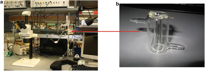
  4. Organ bath, volume 1.2 ml; custom made by glass blower (Fig. ) or large organ baths available commercially.
    Fig 1 a In vitro muscle physiology system and b custom 12 ml organ - photo 1
Next page
Light

Font size:

Reset

Interval:

Bookmark:

Make

Similar books «Skeletal Muscle Regeneration in the Mouse: Methods and Protocols»

Look at similar books to Skeletal Muscle Regeneration in the Mouse: Methods and Protocols. We have selected literature similar in name and meaning in the hope of providing readers with more options to find new, interesting, not yet read works.


Reviews about «Skeletal Muscle Regeneration in the Mouse: Methods and Protocols»

Discussion, reviews of the book Skeletal Muscle Regeneration in the Mouse: Methods and Protocols and just readers' own opinions. Leave your comments, write what you think about the work, its meaning or the main characters. Specify what exactly you liked and what you didn't like, and why you think so.