Case 1
The patient is a 68-year-old white female who was referred for a new evolving rash for 1 week. The patient initially developed red, dime to nickel sized itchy bumps on her back. This rash continued to progress over the next week and eventually involved her bilateral arms, legs, back, abdomen, and face. She complained of an intense itch as well as some associated pain at the lesions that was keeping her awake at night. She had received intramuscular injections of triamcinolone. She stated that this has slowed the formation of new lesions, relieved the tightness in her throat and chest, but there was still an intense itch present. The patient denied any other systemic symptoms present since the start of her rash. The patient was on multiple medicines, including pramipexole, medroxyprogesterone, propranolol, atorvastatin, gabapentin, fenofibrate, aspirin, amlodipine, vitamin B1, vitamin B12, and a multivitamin. The patient had recently been switched from omeprazole to ranitidine approximately 34 days before the first lesion appeared. She has a significant past medical history of squamous cell carcinoma with metastasis to the neck with the treatment of chemotherapy and radiation, chronic bronchitis, hypertension, myocardial infarction, chronic kidney disease, and frequent bladder infections.
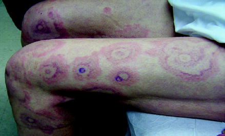
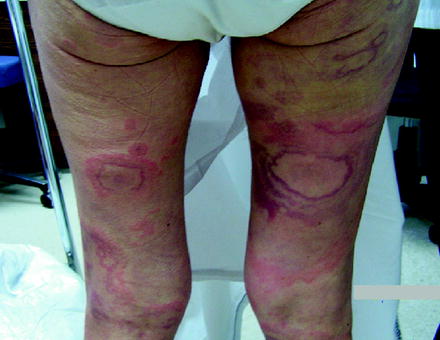
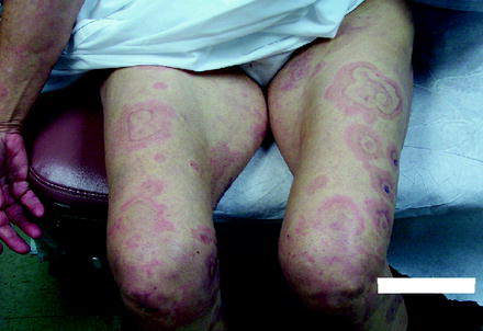
Upon physical examination, the patient demonstrated multiple erythematous, wheal-based, annular to serpiginous, targetoid lesions. The lesions were present on the bilateral legs, arms, back, abdomen, and minimal involvement of the face (Figs. ). No involvement was noted of the mucosal membranes and the patient had no adenopathy present of the cervical, axillary, inguinal, or supraclavicular nodes.
Fig. 1.1
Targetoid, erythematous to violaceous, edematous lesions with central clearing and whealing on the periphery
Fig. 1.2
Targetoid, erythematous to violaceous, edematous lesions with central clearing and whealing on the periphery
Fig. 1.3
Targetoid, erythematous to violaceous, edematous lesions with central clearing and whealing on the periphery
With the Presented Data What Is Your Working Diagnosis?
The clinical presentation of extremely pruritic targetoid urticaria-like lesions that respond to previously given IM steroids most likely suggests some type of urticaria. The tenderness of some of the lesions, the systemic involvement, and the tightness in the throat and chest suggest possibly a vasculitic component. Urticarial vasculitis was favored.
Differential Diagnosis
Acute urticaria, erythema multiforme, urticaria vasculitis, drug eruption, Sweets syndrome, neutrophilic vasculitis.
Workup
A detailed history is imperative in gathering more information once any type of urticaria is suspected. The authors have an urticaria sheet that all patients fill out. This patient had started ranitidine approximately 57 days prior to the development of the first lesion.
A punch biopsy was performed at the center of an active lesion and another at the periphery. The punch biopsy demonstrated a small vessel vasculitis with endothelial swelling and purpura. There was mild focal fibrin deposition, angiocentric inflammatory infiltrate consisting predominantly of neutrophils, and leukocytoclasis.
Serum complement levels (CH50, C3, C4, and anti-C1q) were all within normal levels. Antinuclear antibody (ANA) was negative, and urinary analysis demonstrated moderate amounts of protein. Complete blood count (CBC) and complete metabolic panel (CMP) were unremarkable.
What Is Your Diagnosis and Why?
The diagnosis is urticarial vasculitis secondary to the use of ranitidine. The acute onset of her urticarial breakout after the new use of ranitidine is a large clue to the diagnosis. The punch biopsy is fairly specific for a leukocytoclastic vasculitis consistent with urticarial vasculitis. Urticarial vasculitis has a propensity for eosinophils over other types of vasculitis. Sweetssyndrome demonstrates a more intense dermal infiltrate with marked upper dermal edema. The syndrome typically possesses karyorrhexis and lacks vessel wall necrosis, fibrin, and vasocentric neutrophils, which are features of urticarial vasculitis. The punch biopsy along with clinical features is the standard for diagnosing urticarial vasculitis, but it does not help determine the etiology. Acute urticaria does not demonstrate vasculitic changes.
Management and Follow-Up
The patient was instructed to stop the ranitidine immediately. She was prescribed fexofenadine 180 mg to take daily, hydroxyzine 25 mg at night, and fluocinonide 0.05% cream to apply to the red itchy areas twice a day. The patient was also given pramoxine hydrochloride 1% to apply throughout the day. The lesions completely cleared within 1 week and the proteinuria cleared within 4 weeks. The patient was later rechallenged with the ranitidine and had an acute urticarial dermatitis. Since avoiding ranitidine, the patient has been symptom free.

