• Complain

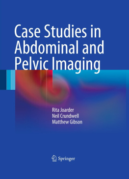
Crundwell Neil - Case Studies in Abdominal and Pelvic Imaging

Here you can read online Crundwell Neil - Case Studies in Abdominal and Pelvic Imaging full text of the book (entire story) in english for free. Download pdf and epub, get meaning, cover and reviews about this ebook. City: London, year: 2011, publisher: Springer-Verlag London Limited, genre: Romance novel. Description of the work, (preface) as well as reviews are available. Best literature library LitArk.com created for fans of good reading and offers a wide selection of genres:

Romance novel Science fiction Adventure Detective Science History Home and family Prose Art Politics Computer Non-fiction Religion Business Children Humor

Choose a favorite category and find really read worthwhile books. Enjoy immersion in the world of imagination, feel the emotions of the characters or learn something new for yourself, make an fascinating discovery.

Crundwell Neil Case Studies in Abdominal and Pelvic Imaging

Case Studies in Abdominal and Pelvic Imaging: summary, description and annotation

We offer to read an annotation, description, summary or preface (depends on what the author of the book "Case Studies in Abdominal and Pelvic Imaging" wrote himself). If you haven't found the necessary information about the book — write in the comments, we will try to find it.

Case Studies in Abdominal and Pelvic Imaging presents 100 case studies, covering both common every-day conditions of the abdomen and pelvis, as well as less common cases that junior doctors and radiologists in training should be aware of. Compiled by experts in the field, Case Studies in Abdominal and Pelvic Imaging uses the most up-to-date and high quality images, including plain films, CT scans, MRI scans and the occasional nuclear medicine image where relevant. Each case is presented in a pedagogical style, with 1-4 images and accompanying questions, followed by answers and further relevant images. This is then augmented by an explanation of the imaging and key teaching points with references for further reading, making this book a valuable learning guide in an accessible form.

Crundwell Neil: author's other books


Who wrote Case Studies in Abdominal and Pelvic Imaging? Find out the surname, the name of the author of the book and a list of all author's works by series.

Case Studies in Abdominal and Pelvic Imaging — read online for free the complete book (whole text) full work

Below is the text of the book, divided by pages. System saving the place of the last page read, allows you to conveniently read the book "Case Studies in Abdominal and Pelvic Imaging" online for free, without having to search again every time where you left off. Put a bookmark, and you can go to the page where you finished reading at any time.

Light

Font size:

Reset

Interval:

Bookmark:

Make
Springer-Verlag London Limited 2011
Rita Joarder , Neil Crundwell and Matthew Gibson Case Studies in Abdominal and Pelvic Imaging 10.1007/978-0-85729-366-4_1
1. Case 1
Rita Joarder 1, Neil Crundwell 1 and Matthew Gibson 2
(1)
Conquest Hospital, St. Leonards-on-Sea, East Sussex, UK
(2)
Royal Berkshire Hospital, Reading, UK
A 78-year-old male with no prior abdominal surgery presented with acute central abdominal pain and vomiting. On examination the abdomen was distended and tympanic with hyperactive bowel sounds. No hernias were palpable. Abdominal x-ray showed dilated small bowel consistent with a distal small bowel obstruction, but cause was not demonstrated.
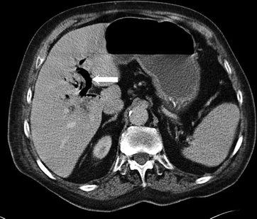
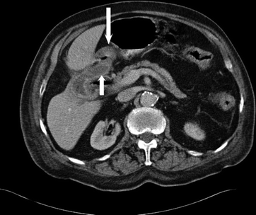
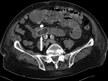
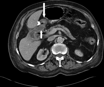
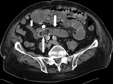
An MDCT of the abdomen and pelvis was performed (Image 1: Axial of upper abdomen, Image 2: Axial of upper abdomen just inferior to Image 1 and Image 3: Axial of lower abdomen).
Image 1 Image 2 Image 3 - photo 1
Image 1
Image 2 Image 3 Questions What is arrowed on Image 1 What is this - photo 2
Image 2
Image 3 Questions What is arrowed on Image 1 What is this sign called - photo 3
Image 3
Questions
What is arrowed on Image 1? What is this sign called?
What are the two organs arrowed on Image 2?
What is arrowed on Image 3? Comment on the small bowel in Image 3.
What is the diagnosis?
Answers
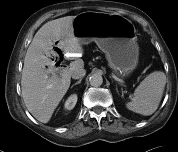
Air in the intrahepatic bile ducts ( arrow Image 4) Aerobilia.
The gallbladder ( short arrow Image 5) and duodenum ( long arrow Image 3) lying closely adjacent with a bubble air ( long arrowhead Image 5) between them.
A gallstone in the ileum ( long arrow Image 6) it has a high density rim with low density material and air within centre of the gallstone. The small bowel proximal to the gallstone is dilated ( medium arrow ) and distal to it is collapsed ( short arrow ), i.e. the gallstone is at the point of transition and the cause of the small bowel obstruction.
Gallstone ileus.
Image 4 Image 5 Image 6 Gallst - photo 4
Image 4
Image 5 Image 6 Gallstone ileus is a rare complication of gallstone - photo 5
Image 5
Image 6 Gallstone ileus is a rare complication of gallstone disease - photo 6
Image 6
Gallstone ileus is a rare complication of gallstone disease, accounting for 14% of all bowel obstructions.
Erosion of a gallstone from the gallbladder to the duodenum causes a biliaryenteric fistula. Cholecystoenteric fistulae occur in less than 1% of patients with gallstones. The gallstone passes through the small bowel and may become impacted causing small bowel obstruction. It is most commonly impacted in the distal ileum but can occur anywhere. An exploratory laparotomy with enterolithotomy is indicated. Enterolithotomy is the most commonly used surgical technique with enterolithotomy combined with cholecystectomy and fistulectomy reserved for selected cases. The clinical presentation depends on impaction site and generally includes abdominal pain, nausea and vomiting. Abdominal x-rays may show the aerobilia, dilated small bowel and sometimes the gallstone; however MDCT is more sensitive and thus the imaging of choice.
Key Points
  • Gallstone ileus is an uncommon cause of small bowel obstruction.
  • Whilst abdominal x-ray may give the diagnosis CT is usually diagnostic.
Further Reading
Ayantunde AA, Agrawal A. (2007) Gallstone ileus: diagnosis and management. World J Surg 31:1292-7 PubMed
Muthukumarasamy G, Venkata SP, Shaikh IA, et al. (2008) Gallstone ileus: surgical strategies and clinical outcome. J Dig Dis 9:156-61 PubMed CrossRef
Springer-Verlag London Limited 2011
Rita Joarder , Neil Crundwell and Matthew Gibson Case Studies in Abdominal and Pelvic Imaging 10.1007/978-0-85729-366-4_2
2. Case 2
Rita Joarder 1, Neil Crundwell 1 and Matthew Gibson 2
(1)
Conquest Hospital, St. Leonards-on-Sea, East Sussex, UK
(2)
Royal Berkshire Hospital, Reading, UK
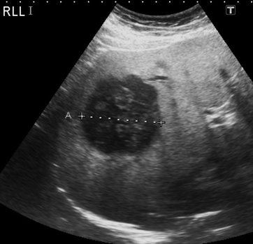
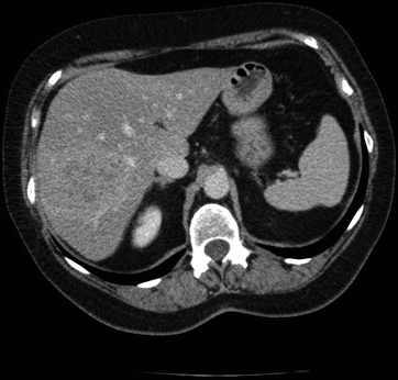
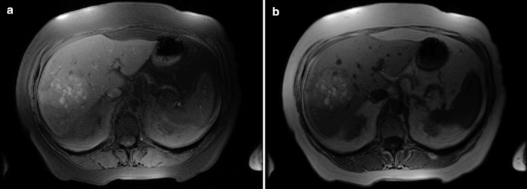
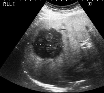
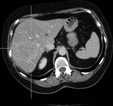
A 70 year-old-female with a false eye complained of right upper quadrant pain. She underwent an US of her upper abdomen (Image 1). An MDCT was then performed (Image 2); this was followed by an MRI of which the axial T1 fat sat and T1 sequences are shown (Images 3a and b).
Image 1 Image 2 Image 3 - photo 7
Image 1
Image 2 Image 3 Questions What are the findings on US What does - photo 8
Image 2
Image 3 Questions What are the findings on US What does the MDCT show - photo 9
Image 3
Questions
What are the findings on US?
What does the MDCT show?
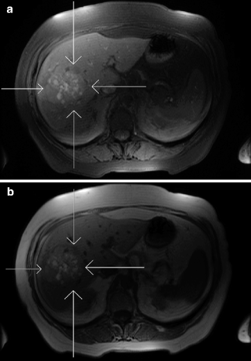
What do the unenhanced T1 fat sat and T1 MR sequences show, and what is their significance?
What is the likely diagnosis?
What further sequences would you perform to clarify appearances?
Answers
The US shows a solitary 5.8 cm solid soft tissue mass within the right lobe of the liver highly suspicious for a metastasis or primary hepatoma (Image 4).
The CT confirms a subtle mass within the right lobe of the liver causing distortion of the adjacent vessels (Image 5).
MRI (often more sensitive than USS) confirms a 6.8 cm mass that is mixed but predominantly high signal on T1 fat sat imaging (Image 6a) and again mixed signal on T1 with focal areas of high signal within it (Image 6b). The areas of high signal on unenhanced T1 fat sat and T1 imaging indicate either haemorrhage or melanin.
The patient had a false eye which would fit with a history of choroidal melanoma, and this is therefore most likely to represent a solitary melanoma metastasis.
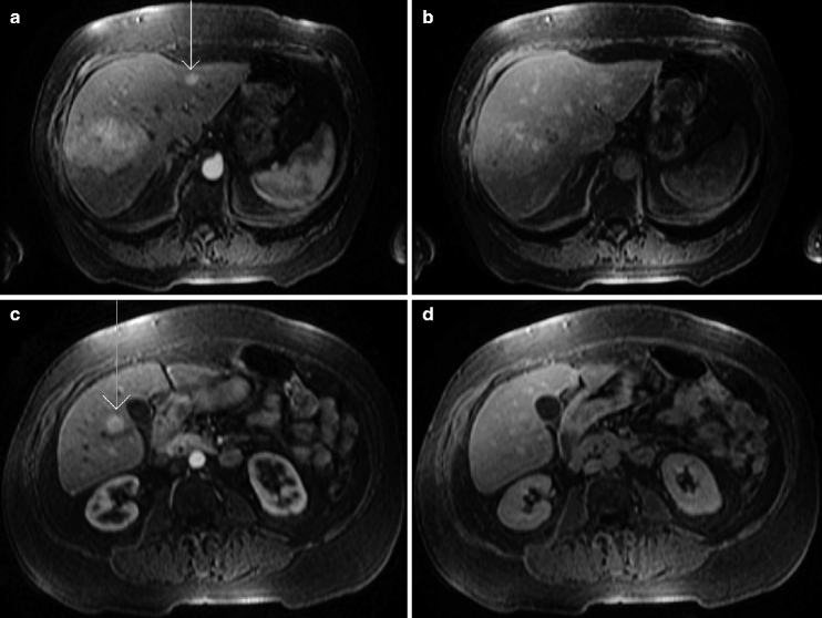
Dynamic post-gadolinium fat sat T1 scans.
Image 4 Image 5 Image 6 - photo 10
Image 4
Image 5 Image 6 Image 7 Dynami - photo 11
Image 5
Image 6 Image 7 Dynamic post-gadolinium fat sat T1 scans Images 7ad - photo 12
Image 6
Image 7 Dynamic post-gadolinium fat sat T1 scans Images 7ad were performed - photo 13
Next page
Light

Font size:

Reset

Interval:

Bookmark:

Make

Similar books «Case Studies in Abdominal and Pelvic Imaging»

Look at similar books to Case Studies in Abdominal and Pelvic Imaging. We have selected literature similar in name and meaning in the hope of providing readers with more options to find new, interesting, not yet read works.


Reviews about «Case Studies in Abdominal and Pelvic Imaging»

Discussion, reviews of the book Case Studies in Abdominal and Pelvic Imaging and just readers' own opinions. Leave your comments, write what you think about the work, its meaning or the main characters. Specify what exactly you liked and what you didn't like, and why you think so.