1.1.1 Image Acquisition and Virtual Rendering of the Patients Head
For proper orthognathic and orthofacial surgery planning, the patients head needs to be scanned without deformation of the facial soft tissue mask, with the mandible in centric relation (CR) and ideally in its individual natural head position (NHP):
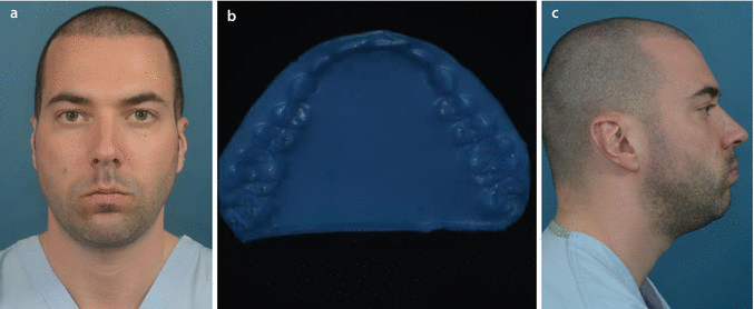
Without deformation of the facial soft tissue mask implements that the patient is scanned in a vertical seated or standing position, without distortion of the facial mask especially lip morphology and posture. With the advent of cone-beam CT imaging (CBCT) in a seated or standing position, this has become feasible. Multi-slice CT (MSCT) scanning, which is performed in a horizontal position and also CBCT apparatus that scan the patient in a supine position, inherently falsify the 3D facial soft tissue mask of the patient due to the effects of gravity. On the other hand, careful attention should be paid that wax-bite wafers or registration devices do not disturb lip position neither lip morphology (Figs. ).
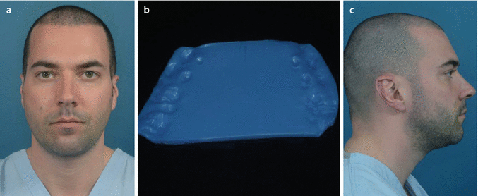
To ensure that the patient is scanned with the mandible in CR, the patient needs to be scanned with a wax-bite wafer. This wax-bite wafer is not different than the one used in conventional orthognathic surgery planning. It is, however, important that this wax-bite wafer is meticulously trimmed in order to avoid deformation of the facial soft tissue mask by interference with the cheeks or lips (Fig. ).
Based on the authors personal experience, the patients head position during image acquisition never corresponds to its true clinical NHP. Therefore, a step-by-step approach is described to virtually modify the patients head position towards its individual clinical NHP (7 see also Sect. ), prior to start 3D virtual treatment planning.
Accurate 3D virtual treatment planning of orthognathic surgery starts with proper image acquisition.
Image Acquisition and Virtual Rendering of the Patients Head
To illustrate the workflow towards (1) image acquisition of the patients head, (2) additional image acquisition of the patients dentition and occlusion and finally (3) additional image acquisition of the texture of the patients head, volunteer M.G. will be used throughout this chapter.
On the other hand, the routine clinical imaging workflow for 3D virtual treatment planning of orthognathic surgery will be demonstrated on Case 1 (Patient V.E.W.) which will be used throughout this book (7 Chaps. ).
Fig. 1.1
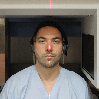
Full face CBCT scanning of the patient, with a wax-bite wafer in CR, needs to be performed in a vertical position (seated or standing) without distortion of the facial soft tissue mask. To avoid movement artefacts, it is crucial that the patient is stabilised by a headrest in the back and a frontal headband. Note that although attempted to scan volunteer M.G. is its individual NHP, in a seated position with the use of laser lines, the shoulders were still distorted which caused a small Yaw rotation (7 see also Sect. ) to the left (i-CAT, Imaging Sciences International Inc., volunteer M.G.)
Stabilisation of the patients head is crucial during CBCT scanning.
Do not use chin supports neither frontal bands that cover the forehead.
Image Acquisition and Virtual Rendering of the Patients Head
Fig. 1.2
A wax-bite wafer (Delar Corp., Lake Oswego, USA) was taken in CR as in conventional treatment planning to ensure adequate CR during CBCT scanning ( b ). Note the significant distortion of the lips and the cheeks when the wax-bite wafer is not trimmed ( a , c ) (volunteer M.G.)
Fig. 1.3
A wax-bite wafer (Delar Corp., Lake Oswego, USA) was taken in CR as in conventional treatment planning to ensure adequate CR during CBCT scanning. The wax-bite wafer was meticulously trimmed to remove all parts that could interfere with the lips and cheeks during CBCT scanning ( b ). Note that there is no distortion of the lips neither of the cheeks ( a , c ) (7 see also Sect. ) (volunteer M.G.)
Image Acquisition and Virtual Rendering of the Patients Head
Fig. 1.4
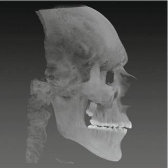
CBCT profile scout view, to ensure that the wax-bite wafer is adequately in place and that the patients mandible is in CR prior to CBCT scanning (i-CAT, Imaging Sciences International Inc., volunteer M.G.)
Always
Take a scout view prior to CBCT scanning of the patient to verify:
Adequate CR
Adequate positioning of the wax-bite wafer
Virtual Rendering of the Patients Head in a 3D Virtual Scene Approach
An appropriate 3D virtual visualisation paradigm (Swennen and Schutyser ) that can be acquired from other 3D imaging sources.
A 3D Virtual Scene Approach is therefore adopted in which the 3D virtual space is considered as a 3D virtual scene with medical image data as actors (Schutyser ). This 3D virtual scene is viewed with a virtual camera and the resulting views are shown on the computer screen. The virtual camera can be moved around in the 3D virtual scene to image and inspect the structures of interest from various angles and positions.
Besides visualising the patients image data with the virtual camera, the 3D virtual visualisation paradigm and the 3D Virtual Scene Approach allow other actions and interactions in the 3D virtual scene such as indicating 3D cephalometric landmarks of the patients hard tissues and teeth (3D-VPS1) (7 see also Sect. ).

