• Complain

Vasken Dilsizian - Atlas of Cardiac Innervation

Here you can read online Vasken Dilsizian - Atlas of Cardiac Innervation full text of the book (entire story) in english for free. Download pdf and epub, get meaning, cover and reviews about this ebook. City: Cham, publisher: Springer International Publishing, genre: Romance novel. Description of the work, (preface) as well as reviews are available. Best literature library LitArk.com created for fans of good reading and offers a wide selection of genres:

Romance novel Science fiction Adventure Detective Science History Home and family Prose Art Politics Computer Non-fiction Religion Business Children Humor

Choose a favorite category and find really read worthwhile books. Enjoy immersion in the world of imagination, feel the emotions of the characters or learn something new for yourself, make an fascinating discovery.

Vasken Dilsizian Atlas of Cardiac Innervation

Atlas of Cardiac Innervation: summary, description and annotation

We offer to read an annotation, description, summary or preface (depends on what the author of the book "Atlas of Cardiac Innervation" wrote himself). If you haven't found the necessary information about the book — write in the comments, we will try to find it.

Vasken Dilsizian: author's other books


Who wrote Atlas of Cardiac Innervation? Find out the surname, the name of the author of the book and a list of all author's works by series.

Atlas of Cardiac Innervation — read online for free the complete book (whole text) full work

Below is the text of the book, divided by pages. System saving the place of the last page read, allows you to conveniently read the book "Atlas of Cardiac Innervation" online for free, without having to search again every time where you left off. Put a bookmark, and you can go to the page where you finished reading at any time.

Light

Font size:

Reset

Interval:

Bookmark:

Make
Springer International Publishing Switzerland 2017
Vasken Dilsizian and Jagat Narula (eds.) Atlas of Cardiac Innervation 10.1007/978-3-319-45800-7_1
1. Anatomy and Molecular Basis of Autonomic Innervation of the Heart
Wengen Chen 1
(1)
Department of Diagnostic Radiology and Nuclear Medicine, University of Maryland School of Medicine, 22 S. Greene Street, Baltimore, MD 21201, USA
Wengen Chen
Email:
Vasken Dilsizian (Corresponding author)
Email:
Keywords
Autonomic nervous system Sympathetic neuronal synapse Signal transduction Reinnervation Molecular regulation
Introduction
The heart is innervated extensively by sympathetic and parasympathetic nerves of the peripheral autonomic nervous system, as well as by sensory nerves. Most sympathetic neurons use norepinephrine as a primary neurotransmitter. Acetylcholine is the main neurotransmitter released by preganglionic and postganglionic vagal nerve terminals, which wrap around the heart and activate the parasympathetic nervous system. Sympathetic and parasympathetic systems exert opposite effects on the heart to regulate the contractile rate and force. Sympathetic nerves increase both heart rate (positive chronotropy) and contractile function (positive inotropy), whereas parasympathetic nerves diminish heart rate (bradycardia) and attenuate sympathetic effects on contractile function. Under normal conditions, the two systems are well balanced to maintain a normal response to external stimulation.
Sympathetic nerves are not distributed homogeneously in the ventricle. The basal inferior region has less innervation compared with the other regions of the left ventricular myocardium []. Maintenance of this physiologic innervation gradient is required to sustain a physiologic repolarization gradient across the ventricular walls. Collapse of the innervation gradient may have an adverse effect on the electrical phenotype of the ventricular wall and cause ventricular arrhythmia.
Several radiotracers have been developed for functional imaging of cardiac sympathetic tone. For example, to target the neurotransmitter norepinephrine, 123I meta -iodobenzylguanidine (123I -m IBG) and 11C-hydroxyephedrine (11C-HED) were developed. Both radiotracers mimic the neuronal transport and storage process of norepinephrine and represent cardiac sympathetic activity. 123I- m IBG is a single-photon emitter and is imaged by using planar or single photon emission computed tomography (SPECT). 11C-HED is a positron emitter and is imaged by using positron emission tomography (PET) or PET/CT hybrid imaging.
Many neuronal factors are involved in sympathetic nerve growth, development, and regeneration after injury, although detailed molecular signaling and regulation mechanisms are not fully understood. Understanding changes and regeneration in sympathetic innervation is more important clinically in the diseased heart than in the normal heart. For example, one of the characteristic neurohormonal changes during heart failure is activation of the sympathetic nervous system. In acute-phase or early heart failure, this activation may preserve cardiovascular homeostasis promptly and even restore cardiac function. However, persistent activation of the sympathetic nervous system in a decompensated heart would be extremely toxic and ultimately would lead to progressively worsening heart function [].
Distribution of Autonomous Nervous System in the Heart
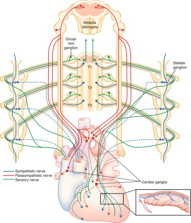
Figure 1-1 Anatomy and distribution of the cardiac nervous systems The - photo 1
Figure 1-1.
Anatomy and distribution of the cardiac nervous systems. The cardiac sympathetic nerve ends ( blue ) are projected from sympathetic neurons in the stellate ganglia, which are bilateral to the thoracic vertebra. The stellate ganglia originate from the spinal cord (dorsal root ganglia or preganglia). The cardiac parasympathetic nerves ( red ) extend from parasympathetic neurons in the cardiac ganglia, which are located at the base of both atria, and are direct branches of the vagus nerve in the human from the brainstem (10th cranial verve). The sensory nerves ( green ) project to the upper thoracic dorsal horn via dorsal root ganglia, which are responsible for pain perception and initiation of a protective response during cardiac injury. The inset indicates the transverse section of the heart and demonstrates the distribution of sympathetic nerve fibers within the left ventricle. The sympathetic nerve fibers run in the epicardial area and then individually dive into the myocardial wall, and display an epicardial-to-endocardial gradient in the heart []).
Sympathetic Neuronal Synapse in the Heart
Figure 1-2 Sympathetic neuronal synapse in the heart The sympathetic neuronal - photo 2
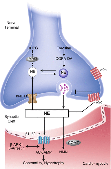
Figure 1-2.
Sympathetic neuronal synapse in the heart. The sympathetic neuronal transmitter norepinephrine ( NE ) is synthesized from tyrosine. First, it is converted to dopamine by cytoplasmic enzymes. Dopamine then is transported to intraneuronal vesicles, where NE is synthesized and stored. Upon neuronal stimulation, the vesicles fuse with the presynaptic membrane and release the NE into the cleft. The released NE then binds to the postsynaptic - and -adrenergic receptors, which are G-proteincoupled receptors, to transduce signals to improve excitationcontraction coupling of the myocardium and to increase heart rate and force. Cleft NE also activates the presynaptic 2-adrenergic receptors to inhibit further NE release. The effect of the NE is terminated by two mechanisms: approximately 80 % of the NE is taken up to the presynaptic area by an ATP-dependent active process via a transporter called human norepinephrine transporter 1 (hNET1; uptake-1 system), whereas the reminder spills over into the nonneuronal tissue and bloodstream (uptake-2 system). Thus, several molecules in this pathway may serve as targets for the functional imaging of sympathetic tone in the heart. For example, to target the neurotransmitter NE, 123I -m IBG and 11C-HED were developed. Both radiotracers mimic the neuronal transport and storage process of NE and represent cardiac sympathetic activity. hNET1 also may be an ideal target to represent the uptake-1 system activity in the heart. AC adenyl cyclase, AMP adenosine monophosphate, cAMP cyclic adenosine monophosphate, COMT catechol- O -methyltransferase, DA dopamine, DHPG dihydroxyphenylglycol, DOPA dihydroxyphenylalanine, MAO monoamine oxidase, NMN normetanephrine (Data from Haider et al. []).
Figure 1-3 Down-regulation of the presynaptic hNET1 in heart failure In - photo 3
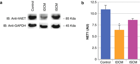
Figure 1-3.
Down-regulation of the presynaptic hNET1 in heart failure. In patients with heart failure (ischemic or nonischemic), initially there is increased sympathetic activity with increased release of NE from the presynaptic vesicles as a compensatory reaction to contribute to increased cardiac contractility. ( a , b ) There is corresponding down-regulation of the hNET1 transporter (decreased uptake-1 system) in idiopathic dilated cardiomyopathy (IDCM) and ischemic cardiomyopathy (ISCM) []).
Signal Transduction of the Postsynaptic Cardiac Adrenergic Receptors
The human genome encodes nine different adrenoceptor genes. They are grouped into three G-proteincoupled receptor (GPCR) families with three members each, namely the 1-adrenoceptors (1A, 1B, and 1D), 2-adrenoceptors (2A, 2B, and 2C), and -adrenoceptors (1,2, and 3) []. The 1 subtype mediates an increase in cardiac contractility, whereas the 2 subtype is responsible for vasodilatation. These receptors are not just signal transducers but also components of a complex and highly regulated signaling machinery. Adrenoceptors consist of seven transmembrane (TM)-spanning -helical domains, an extracellular region (N terminus, three extracellular loops), and an intracellular region (three intracellular loops, C terminus).
Next page
Light

Font size:

Reset

Interval:

Bookmark:

Make

Similar books «Atlas of Cardiac Innervation»

Look at similar books to Atlas of Cardiac Innervation. We have selected literature similar in name and meaning in the hope of providing readers with more options to find new, interesting, not yet read works.


Reviews about «Atlas of Cardiac Innervation»

Discussion, reviews of the book Atlas of Cardiac Innervation and just readers' own opinions. Leave your comments, write what you think about the work, its meaning or the main characters. Specify what exactly you liked and what you didn't like, and why you think so.