1. Ins and Outs of the Cardiac Chambers
The flow of blood through the normal, healthy heart is always unidirectional, in both the right and left sides of the heart. This is endured by the sequential, opening and closing of the atrioventricular valves and the aortic/pulmonary valves. Blood flows when there is a pressure gradient. The phasic changes in atrial and ventricular pressure during a cardiac cycle determine, in concert with gating valves, the unidirectional intracardiac flow. The student should understand what generates the pressure that ejects blood volume from each ventricle and what pressure gradient drives the inflow or ventricular infilling of blood during diastolic relaxation phase of the cardiac cycle. The questions below address the physiology of some of these cardiac events.
1. What cardiac index is used as a quantitative measure of myocardial contraction strength?
Answer
Myocardial contractility is the term for the power of cardiac muscle contraction and is represented by the ejection fraction.
Concept
Cardiac muscle contract as for skeletal muscles. Both muscle types perform work, the skeletal muscles in isotonic contraction and the heart does cardiac work in ensuring a continuous blood perfusion to all the peripheral tissues. The strength of cardiac muscle contraction can also be increased. In the skeletal muscles, graded muscle tension is increased by recruitment of more motor units and higher frequency of motor nerve impulses to produce summative, titanic contraction.
In cardiac muscles, the strength is increased by cardiac sympathetic nerve and circulating hormones, the main one being adrenaline, that binds to beta adrenergic receptors on the cardiac muscles, that are also activated by neurotransmitter noradrenaline released from the sympathetic fibers.
The increased contractility is also represented by the increased ejection fraction. The ejection fraction is the ratio of the ejected stroke volume and the end-diastolic volume (EDV) in the ventricle before contraction. For a given EDV, more volume is pumped out by the more contractile ventricle. The volume remaining in the ventricle after systolic contraction, the end-systolic volume will be reduced when myocardial contractility is increased.
In hyperthyroidism, the contractility is also increased by the excess circulating thyroid hormones. Thyroid hormones upregulate beta adrenergic receptors on the cardiac muscle and potentiates the sympathetic/adrenaline positive inotropic actions. Positive inotropism means the same as increased myocardial contractility/higher ejection fraction. Thyroid hormones can also alter the myosin ATPase type in the cardiac muscle, which also accounts for the greater contractility.
2. In Starlings mechanism of the heart, what are the y - and x -axes of the Starlings graph?
Answer
The x -axis is the EDV and the y -axis is the stroke volume.
Concept
The mechanical property of the cardiac ventricle muscles described by Starling is an intrinsic muscle phenomenon. By intrinsic this means that no extrinsic nerve or circulating hormones play a role in the Starlings mechanism (or Law) of the heart.
The heart is a generous organ. If it receives more blood volume, it will give out more blood volume. The heart does not hoard! Using cardiac volumes to describe Starlings event, this states that the greater the physiologic increase in the EDV, the bigger will be the stroke volume.
The axes of the graph can also represent x -axis as ventricular filling (venous return) and y -axis as cardiac output. A larger EDV stretches the ventricular muscle and the contracting tension is greater. The histophysiologic basis for this is the degree of potential overlap between the actin and myosin filaments in the cardiac muscle at different lengths.
Up to an optimal length, the increase in EDV and hence cardiac muscle length will be followed by a larger ejected, stroke volume.
Note that the ejection fraction is unchanged (this fraction is a measure of myocardial contractility, question 1 above).
This more in more out ventricular Starlings mechanics applies in both the left and right ventricles. The maximum systolic intraventricular pressure is much higher in the left ventricle (120 mmHg) compared to that in the right ventricle (~ 30 mmHg). However, the stroke volume (SV) of each ventricle is the same, because the cardiac work has to be higher for the left ventricle against a higher afterload (~ 100 mmHg) than the afterload at the right side represented by the mean pulmonary arterial pressure.
The intraventricular and aortic/pulmonary vascular pressures are different on each side of the heart, but the volume dynamics (EDV and SV) of the Starlings cardiac mechanism is the same and operative in both ventricles.
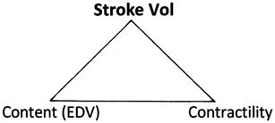
The ejected blood volume with each heartbeat (stroke volume, SV) is determined by two contributing factors. One is an intrinsic cardiac muscle mechanism (Starlings law) where SV is dependent on ventricular filling ( EDV ). The other way to increase SV is by an increased cardiac sympathetic nerve action or by higher circulating adrenaline that both produce a greater myocardial contractility. Increased contractility is defined by a bigger ejection fraction (SV/EDV)
3. What mechanism ensures that the right and left ventricular cardiac outputs are equalized over time?
Answer
Starlings mechanism of the heart has the essential physiologic role in equalization the cardiac output of the two ventricular pumps that are arranged in series.
Concept
The figure in physiology texts sometime gives the students the impressions that the systemic and the pulmonary circulations are in parallel. If we imagine stretching out the whole circulatory system into a chain, the right and the left ventricles would be seen to be connected in series like two beads along the bloody vascular chain.
The serial arrangement of the right and left ventricular pumps present a potential problem for the vascular blood traffic flow. It is crucial that the two pumps are synchronized with regards to the cardiac output put out be each ventricle. Should there be unequal cardiac outputs, very soon we will have problems of vascular traffic congestion.
Beat by beat, there could be small fluctuations in the stroke volume from each ventricle. There are 60 beats in each minute, and the differences in the stroke volumes will accumulate to produce unequal cardiac output if there is no mechanism to adjust for this.
This is where the intrinsic Starlings myocardial mechanics becomes important. If one ventricle has a larger cardiac output, this will mean a greater filling of the other ventricle. The second ventricle then contracts more strongly. If the second ventricle does not intrinsically pumps out more of what it has received (increased EDV), then the traffic upstream from the second ventricle will be congested.
To give a clinical illustration, if the right ventricle weakens, there will be venous congestion (with development of peripheral edema). On the other hand (heart), left ventricular failure will result in pulmonary venous congestion and this can cause pulmonary edema.

