RADIOLOGY OF
AUSTRALIAN
MAMMALS
LARRY VOGELNEST AND GRAEME ALLAN

Larry Vogelnest and Graeme Allan 2015
All rights reserved. Except under the conditions described in the Australian Copyright Act 1968 and subsequent
amendments, no part of this publication may be reproduced, stored in a retrieval system or transmitted in any form or by
any means, electronic, mechanical, photocopying, recording, duplicating or otherwise, without the prior permission of the
copyright owner. Contact CSIRO Publishing for all permission requests.
National Library of Australia Cataloguing-in-Publication entry
Vogelnest, L. (Larry), author.
Radiology of Australian mammals / Larry Vogelnest and Graeme Allan.
9780643108646 (hardback)
9780643108653 (epdf)
9780643108660 (epub)
Includes bibliographical references and index.
Mammals Australia.
Veterinary radiology.
Allan, Graeme Sutcliffe, author.
599.0994
Published by
CSIRO Publishing
Locked Bag 10
Clayton South VIC 3169
Australia
Telephone: +61 3 9545 8400
Email:
Website: www.publish.csiro.au
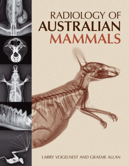
Front cover (main image): tammar wallaby (Macropus eugenii)
Front cover (top to bottom): yellow-bellied glider (Petaurus australis), common wombat (Vombatus ursinus), platypus
(Ornithorhynchus anatinus), sub-adult eastern grey kangaroo (Macropus giganteus), ghost bat (Macroderma gigas)
Back cover (top to bottom): squirrel glider (Petaurus norfolcensis), common wombat (Vombatus ursinus), spot-tailed quoll
(Dasyurus maculatus), spot-tailed quoll (Dasyurus maculatus), greater bilby (Macrotis lagotis)
Set in 10.5/14 Palatino & Optima
Edited by Kerry Brown
Cover design by James Kelly
Typeset by Thomson Digital
Index by Bruce Gillespie
Printed in China by 1010 Printing International Ltd
CSIRO Publishing publishes and distributes scientific, technical and health science books, magazines and journals from
Australia to a worldwide audience and conducts these activities autonomously from the research activities of the
Commonwealth Scientific and Industrial Research Organisation (CSIRO). The views expressed in this publication are
those of the author(s) and do not necessarily represent those of, and should not be attributed to, the publisher or CSIRO.
The copyright owner shall not be liable for technical or other errors or omissions contained herein. The reader/user
accepts all risks and responsibility for losses, damages, costs and other consequences resulting directly or indirectly from
using this information.
Original print edition:
The paper this book is printed on is in accordance with the
rules of the Forest Stewardship Council. The FSC promotes
environmentally responsible, socially beneficial and
economically viable management of the worlds forests.
CONTENTS
Larry Vogelnest, Graeme Allan and Susan Hemsley
Nadine Fiani
Frances Hulst, Graeme Allan and Larry Vogelnest
Paul Andrew
This text has been compiled to fill a void in the current literature. We recall on many occasions, while interpreting radiographs of various Australian mammals, saying We need to do a text on the normal radioanatomy of Australian mammals. We have acted on it and produced Radiology of Australian Mammals.
The recognition, diagnosis and treatment of injury and disease in wildlife species present unique challenges for the veterinarian. This is particularly true for many species of Australian native mammals. Many are cryptic in nature and frequently mask their clinical signs when injured or suffering from disease the preservation response. When animals can no longer compensate and appear clinically affected, the effects of their injuries or diseases are advanced and efforts to treat these animals are often fruitless. Veterinarians dealing with wildlife must draw on all their skills and powers of observation to recognise the signs of injury and disease. In the majority of cases, further examination and diagnostic workup will be required to confirm a diagnosis. As with domestic species, a suite of diagnostic modalities can be used to further define the nature and extent of injury or disease, guide therapeutic decisions and determine prognosis. Radiology is a fundamental diagnostic modality that is generally readily available and, with the advent of digital radiology, provides high-quality images that can be viewed almost instantly. These can also be easily electronically transferred for referral to a specialist wildlife veterinarian or radiologist for interpretation and opinion if required.
Two important aspects of radiology are the recognition and description of abnormal findings and the interpretation of these findings. In order to recognise abnormalities, knowledge of normal radioanatomy is required. This is particularly challenging with wildlife because of the frequent lack of familiarity with a species and anatomic descriptions, and because radiographic images of normal animals are not readily available for comparison. A further challenge is the variation in anatomy among the different wildlife species; in some instances, even those within the same taxonomic group. One option that can increase ones confidence in distinguishing normal from abnormal is consultation of reference sources on normal radioanatomy. This, until now, has not been available for Australian mammals. Radiology of Australian Mammals is the first comprehensive resource on the normal radioanatomy of Australian mammals.
The primary aim of this text is to provide a detailed reference on the normal radioanatomy of Australian mammals. As there are over 380 native mammal species in 48 families and 150 genera in Australia, to represent all of these radiographically would be impossible. We have used selected, readily available species to represent a typical species within each of the major taxonomic groups. We have not included rodents, as Australian native rodents are not anatomically sufficiently different from exotic rodent species described in other texts. We have not included marine mammals, as acquiring complete image sets for available species was not possible. We have also not included the dingo, as this species has similar radioanatomy to that of domestic canids. In most cases clinically normal, healthy animals were used. In some instances, radiographs were opportunistically obtained from animals undergoing investigative procedures for injury or disease. After careful scrutiny by the authors, only radiographs representing normal radioanatomy have been used in Chapters 2 to 9. In some cases, however, radiographs with abnormalities were selected if the abnormality did not detract from the intent to demonstrate other normal radiographic features. Radiographic abnormalities in these images are indicated by annotations on the images and described in the caption. Variations of normal radioanatomy, for example between sexes or ages, have been indicated where relevant. The animals used were primarily sourced from the animal population at Taronga Zoo or wildlife admitted to the Taronga Wildlife Hospital, Sydney, Australia. Animal use was approved by the Zoos Animal Ethics Committee (approval numbers 4a/11/10 and 4a/10/13). All animals were anaesthetised for radiographic studies and strict radiation safety protocols were adhered to for personnel. A small number of cadaver specimens were also used. If species or specific images were not available from Taronga, these were sourced from other institutions and have been acknowledged appropriately. The majority of images are conventional digital radiographs. A small selection of computed tomography images is included. No magnetic resonance images have been used. A chapter on radiographic technique is included and provides useful information on radiography of wildlife species, highlighting the unique challenges inherent in working with wildlife. There are nine chapters covering the major Australian mammal taxonomic groups. Each chapter covers radioanatomy of the skeletal system and major soft tissue structures and systems. A chapter on dental radiology discusses and demonstrates normal dental radioanatomy of Australian marsupials and monotremes. The final chapter includes selected radiographic pathology case studies, providing an appreciation of radiographic findings seen in some common or noteworthy diseases of Australian mammals. Anatomical descriptions are based on available literature (some of which is antiquated), examination of skeletal and cadaver specimens and comparison with domestic species anatomy. There were many gaps in anatomical descriptions and in some instances skeletal structures identified on radiographs had not previously been described.
Next page
