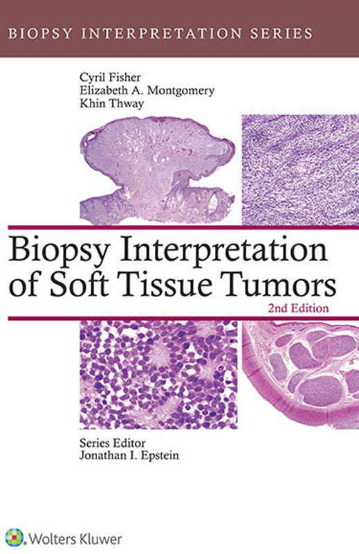
Elizabeth Montgomery - Biopsy Interpretation of Soft Tissue Tumors
Here you can read online Elizabeth Montgomery - Biopsy Interpretation of Soft Tissue Tumors full text of the book (entire story) in english for free. Download pdf and epub, get meaning, cover and reviews about this ebook. year: 2015, publisher: LWW, genre: Science. Description of the work, (preface) as well as reviews are available. Best literature library LitArk.com created for fans of good reading and offers a wide selection of genres:
Romance novel
Science fiction
Adventure
Detective
Science
History
Home and family
Prose
Art
Politics
Computer
Non-fiction
Religion
Business
Children
Humor
Choose a favorite category and find really read worthwhile books. Enjoy immersion in the world of imagination, feel the emotions of the characters or learn something new for yourself, make an fascinating discovery.

- Book:Biopsy Interpretation of Soft Tissue Tumors
- Author:
- Publisher:LWW
- Genre:
- Year:2015
- Rating:5 / 5
- Favourites:Add to favourites
- Your mark:
Biopsy Interpretation of Soft Tissue Tumors: summary, description and annotation
We offer to read an annotation, description, summary or preface (depends on what the author of the book "Biopsy Interpretation of Soft Tissue Tumors" wrote himself). If you haven't found the necessary information about the book — write in the comments, we will try to find it.
Key Features:
Stay up to date with whats new in soft tissue pathology with coverage of the many recent advances in immunohistochemistry and discoveries of underlying molecular events.
Clearly visualize the diagnostic challenges you face with more than 300 full-color, high-quality images in print, and over 1,400 more online.
Focus on differential diagnosis with numerous tables of specific morphologic features and use of immunohistochemical panels.
See the complete picture, including relevant clinical and management points impacted by the pathologic diagnosis, and updated literature that includes new entities and references.
Test your knowledge online with 50 multiple-choice questions ideal for self-assessment.
Now with the print edition, enjoy the bundled interactive eBook edition, which can be downloaded to your tablet and smartphone or accessed online and includes features like:
Complete content with enhanced navigation
Powerful search tools and smart navigation cross-links that pull results from content in the book, your notes, and even the web
Cross-linked pages, references, and more for easy navigation
Highlighting tool for easier reference of key content throughout the text
Ability to take and share notes with friends and colleagues
Quick reference tabbing to save your favorite content for future use
Elizabeth Montgomery: author's other books
Who wrote Biopsy Interpretation of Soft Tissue Tumors? Find out the surname, the name of the author of the book and a list of all author's works by series.


Biopsy Interpretation of Soft Tissue Tumors — read online for free the complete book (whole text) full work
Below is the text of the book, divided by pages. System saving the place of the last page read, allows you to conveniently read the book "Biopsy Interpretation of Soft Tissue Tumors" online for free, without having to search again every time where you left off. Put a bookmark, and you can go to the page where you finished reading at any time.
Font size:
Interval:
Bookmark:

INTRODUCTION
Soft tissue tumors comprise a group of entities showing mesenchymal differentiation which can be located in skin, subcutis, or deep soft tissue. The latter includes subfascial limb and limb girdle tumors and those located in head and neck, abdomen, retroperitoneum (including paratesticular region), pelvis, and thoracic and intracranial cavities. Similar lesions can also involve viscera. Typically, soft tissue tumors are classified by the type of differentiation which they display, and the principal objectives of the surgical pathologist are to identify the lineage of the lesional cells and assess their malignant potential. Nonmesenchymal lesions such as carcinoma or melanoma can also present as soft tissue neoplasms.
In many cases, the tumor is seen initially as a spindle cell, epithelioid cell, small round cell, or pleomorphic lesion that needs to be characterized further with the aid of ancillary techniques including immunohistochemistry and molecular pathologic analyses. Lesions with adipose, osteochondroid, or vascular space formation can be identified morphologically but often present difficulties in precise subcategorization and in assessment of malignancy. This book is, therefore, organized into chapters representing the main morphologic categories as they present to the pathologist. Each entry includes relevant clinical data, morphologic features, and information from ancillary techniques wherever appropriate. The differential diagnosis within each category is presented in detailed tabular form. It is hoped that this approach will reflect the pathologists diagnostic process and will facilitate diagnosis and provision of relevant information to the clinician in this rare and difficult group of neoplasms. Necessarily, some entities appear in more than one category, with the main entry corresponding to the most common pattern or location in each case.
BIOPSY
Most superficial soft tissue tumors can be directly excised in their entirety, but deep tumors often grow to a large size and require diagnosis by biopsy. Interpretation can be difficult because a biopsy represents only a very small proportion of the tumor and the appearances in sarcomas can be heterogeneous and modified by myxoid, fibrous, or inflammatory stromal changes. Accurate diagnosis involves two major decision making steps: Is the lesion malignant? If so, what sort of a sarcoma is it? Tumor grade should also be assessed when possible. Because of the rarity of soft tissue tumors, a patient with a soft tissue mass should ideally be referred to a specialized center for evaluation, biopsy (or review of pathology if carried out elsewhere), and, if necessary, treatment by a multidisciplinary team that includes an experienced pathologist.
Biopsy Techniques
Biopsy of a soft tissue tumor can be excisional, open incisional, or closed.
Excisional biopsy aims to remove the whole lesion for both diagnostic and therapeutic purposes and is usually indicated for superficial lesions and those <5 cm in diameter. The excision should include a margin of normal tissue, although microscopic examination might later reveal tumor at the margin, indicating further excision. This applies especially to lesions that are shelled out as they are generally removed through the false capsule within which tumor cells remain.
Open incisional biopsy involves surgical opening of skin and subcutaneous tissues and direct sampling of the tumor from which a wedge can be removed. This approach results in a larger amount of tissue for diagnosis but has the potential complications of a formal surgical procedure, including wound dehiscence and infection, and compromise of subsequent definitive surgery or radiation therapy. It can also facilitate spread of tumor from the main lesion into adjacent or overlying tissues. In addition, an open procedure also requires more resources and is therefore less cost-effective than closed methods.
Closed biopsies carry minimal risk of infection or seeding of the wound with tumor and provide adequate material for all investigative modalities. They are simple and relatively inexpensive office procedures that are readily repeated if more material is required. Techniques include core needle biopsy (CNB) and fine needle aspiration cytology (FNAC).
(a) CNBs are usually taken with a Tru-Cut needle (CareFusion Corp., San Diego, CA), which can be inserted through a tiny skin incision and angled to sample different parts of the tumor. Several studies of cutting needle biopsies for soft tissue tumors have demonstrated that the larger the needle, the more optimal the specimen. Ultrasound- or computerized axial tomographyguided biopsy can be performed for less accessible lesions such as those deep in body cavities, although they usually yield thinner cores because of the use of a smaller gauge needle. The use of CNB has become more widespread in recent years, and practiced operators can achieve a very high proportion of adequate biopsies. An experienced pathologist can differentiate benign from malignant tumors with a sensitivity of 99.4%, a specificity of 98.7%, a positive predictive value of 99.4%, and a negative predictive value of 98.7%. Ninety-five percent accuracy can be obtained in subtyping and 85% in grading of soft tissue neoplasms on needle biopsies. Grading is less accurate than histologic typing, usually because a higher grade area is identified in the excised specimen. However, an at least grade, or an indication of low- or high-grade malignancy, can be given. For some pediatric tumors, grading, classification, and prognostic categorization have specific issues that may limit the use of CNBs.
(b) FNAC is used for initial diagnosis of soft tissue tumors in some large centers and gives good results in expert hands. FNAC can have a role, however, in identifying recurrent or metastatic sarcomas.
Following prior biopsy diagnosis, frozen section is rarely indicated for immediate management, as margins are better assessed on the excised specimen. Intraoperative pathologic examination is, however, sometimes required at open biopsy, either for diagnosis or to confirm that adequate viable tissue is available.
SPECIMEN COLLECTION AND HANDLING
Core Biopsies
A minimum of three needle biopsy cores is optimal for morphology, immunohistochemistry, touch imprints and molecular genetic analysis (which may be done on paraffin sections), cytogenetics, and storage of frozen tissue for future molecular studies. Specimen adequacy can be evaluated with touch imprints or frozen section at the time of the biopsy procedure. Touch imprints are useful for a variety of possible genetic and cell marker studies, and some experts have advocated performing touch imprints until the pathologist is comfortable with the adequacy of the entire specimen. This is not, however, usually necessary in routine use. If required, one core can be used for frozen section examination and a portion can be placed in glutaraldehyde for possible ultrastructural studies. The remainder of the cores should be submitted in formalin.
CNBs should be counted and measured. If the cores are not fragmented, and resources allow, it is preferable to separate and embed them in more than one cassette to allow maximal use of the available material.
Open Biopsies
These should also be submitted in formalin, but if the specimen is large enough, a small portion can be frozen in liquid nitrogen and stored at 80C.
Resection Specimens
Surgical excision specimens should be sent to the laboratory directly after removal from the patient, without formalin, to allow orientation and sampling of fresh tissue for genetic studies. The latter might include freezing a suitable portion in liquid nitrogen and storage at 80C in an appropriate facility. All likely investigative options should be considered at the time that fresh tissue arrives in the laboratory to maximize potential diagnostic and prognostic information.
Font size:
Interval:
Bookmark:
Similar books «Biopsy Interpretation of Soft Tissue Tumors»
Look at similar books to Biopsy Interpretation of Soft Tissue Tumors. We have selected literature similar in name and meaning in the hope of providing readers with more options to find new, interesting, not yet read works.
Discussion, reviews of the book Biopsy Interpretation of Soft Tissue Tumors and just readers' own opinions. Leave your comments, write what you think about the work, its meaning or the main characters. Specify what exactly you liked and what you didn't like, and why you think so.