Overview
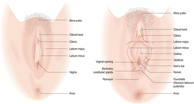
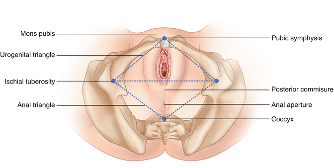
The vulva consists of the female genital structures external to the vaginal openingthe introitus. Anatomically, the vulva lies within the perineum, which is a diamond-shaped region bounded anteriorly by the pubic symphysis, laterally by the left and right ischial tuberosities of the pelvic bones, and posteriorly by the coccyx (Figs. ]. The perineum further subdivides into an anterior urogenital triangle with the vulva and a posterior anal triangle with the anus and its external sphincter. The vulva comprises the mons pubis, the labia majora, the labia minora, the clitoris, and the vestibule. The vestibule itself is the specific region demarcated anteriorly by the clitoral prepuce, laterally by the labia minora, and posteriorly by the fourchette, which is a fold of skin where the labia minora join. Within this region are the clitoris, the vestibulovaginal bulbs and associated vestibular (Bartholins) glands, the urethral meatus and associated periurethral (Skenes) glands, and the vaginal introitus. Before considering the anatomy and histology of these structures in more detail, however, it will be beneficial to first consider the development of the female reproductive tract.
Fig. 1.1
External genitalia of the vulva (Used with permission from Robboy et al. []
Fig. 1.2
Perineal surface anatomy
Embryology of the Female Reproductive Tract and External Genitalia
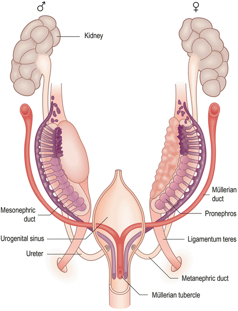
The urinary and reproductive systems in both males and females are embryologically and anatomically interrelated in that both develop from a urogenital ridge of intermediate mesoderm located along the posterior body wall in the developing abdominal cavity and both open into an endoderm-lined cloaca at the caudal end of the embryo (Table ]. The excretory tubules elongate into S-shaped loops encapsulating a rudimentary glomerular capillary tuft located along their medial portion. The lateral end of each excretory tubule attaches to a collecting duct running longitudinally known as the mesonephric or Wolffian duct, which opens into a portion of the cloaca that will invaginate to form the urogenital sinus. As the name implies, the urogenital sinus contributes to the lower urinary tract, namely, the bladder and urethra, as well as a portion of the reproductive tracts in both the male (prostate and prostatic urethra) and female (vagina and vestibule). Initially, the segmental nephrons of the mesonephros provide functional urine output but then regress as the definitive, metanephric kidneys arise from the caudal intermediate mesoderm. The longitudinal mesonephric duct on each side persists and becomes part of a paired set of genital ducts that contribute significantly to the male reproductive tract. As described below, the mesonephric ducts largely regress in the female and normally only contribute to rudimentary structures.
Table 1.1
Homologues and origins of the human reproductive system
Indifferent | Germ layer | Female | Male |
|---|
Gonad | Mesoderm | Ovary | Testis |
Paramesonephric (Mllerian) duct | Mesoderm | Fallopian tubes | Appendix testis |
Paramesonephric duct | Mesoderm | Uterus, vagina | Prostatic utricle |
Mesonephric (Wolffian) duct | Mesoderm | Rete ovarii | Rete testis |
Urogenital sinus | Endoderm | Skenes glands | Prostate |
Urogenital sinus | Endoderm | Bladder, urethra | Bladder, urethra |
Urogenital sinus | Endoderm | Bartholins gland | Bulbourethral gland |
Labioscrotal folds | Ectoderm | Labia majora | Scrotum |
Urogenital folds | Mesoderm | Labia minora | Spongy urethra |
Genital tubercle | Mixed | Vestibular bulbs | Bulb of the penis |
Genital tubercle | Mixed | Clitoral glans | Glans penis |
Genital tubercle | Mixed | Clitoral crura | Crus of the penis |
Prepuce | Mixed | Clitoral hood | Foreskin |
Gubernaculum | Mesoderm | Round ligament of the uterus | Gubernaculum testis |
Modified from Ref. []
Fig. 1.3
Anlage of the genital organs in the indifferent, bisexual stage (Used with permission from Jaubert et al. []
During the 6th week, primordial germ cells appear within the medial, or genital, portion of the urogenital ridge. These germ cells initially arise in the embryos epiblast, migrate through the primitive streak during gastrulation in the 3rd week, and come to rest in the wall of the yolk sac near the forming allantois. Soon after gastrulation, the germ cells migrate back into the embryo along the dorsal mesentery of the hindgut and invade the medial edge of the urogenital ridge. In response to the germ cells, the overlying coelomic epithelium of the genital ridge proliferates and invades the mesenchymal tissue to form a series of irregularly shaped, primitive sex cords that remain connected to the surface epithelium and become closely associated with the germ cells. The surrounding mesenchymal cells, in turn, will develop into sex-specific interstitial cells of the gonads that contribute to the differentiation of the male or female phenotype.
During this same period, a second set of genital ducts, the paramesonephric (Mllerian) ducts, arise as the epithelium along the lateral edge of the genital ridge adjacent to the mesonephric ducts invaginates to form a longitudinal tube. At their cranial end, the paramesonephric ducts are lateral to the mesonephric ducts and end in a funnel-shaped opening into the abdominal cavity at about the same level as the superior aspect of the indifferent gonad. Caudally, the paramesonephric ducts continue lateral to the mesonephric ducts and then cross under (ventral to) the mesonephric ducts to course medially and partially fuse in the midline to form the uterine canal. The uterine canal projects caudally until it meets the wall of the urogenital sinus, where it causes a small swelling known as the paramesonephric, or Mllerian, tubercle to form [].

