• Complain

Laurence Brown (editor) - Pathology of the Vulva and Vagina

Here you can read online Laurence Brown (editor) - Pathology of the Vulva and Vagina full text of the book (entire story) in english for free. Download pdf and epub, get meaning, cover and reviews about this ebook. year: 2012, publisher: Springer Nature, genre: Romance novel. Description of the work, (preface) as well as reviews are available. Best literature library LitArk.com created for fans of good reading and offers a wide selection of genres:

Romance novel Science fiction Adventure Detective Science History Home and family Prose Art Politics Computer Non-fiction Religion Business Children Humor

Choose a favorite category and find really read worthwhile books. Enjoy immersion in the world of imagination, feel the emotions of the characters or learn something new for yourself, make an fascinating discovery.

Laurence Brown (editor) Pathology of the Vulva and Vagina

Pathology of the Vulva and Vagina: summary, description and annotation

We offer to read an annotation, description, summary or preface (depends on what the author of the book "Pathology of the Vulva and Vagina" wrote himself). If you haven't found the necessary information about the book — write in the comments, we will try to find it.

Doctors and the general public are increasingly recognising diseases of the vulva and vagina as a cause of sexual dysfunction, morbidity and death, yet the wide but sometimes rare range of conditions involving this area are poorly represented in most textbooks of pathology. As the first volume in the Essentials of Diagnostic Gynecological Pathology series sponsored by the British Association of Gynecological Pathologists, Pathology of the Vulva and Vagina is one of the very few dealing wholly with this subject.

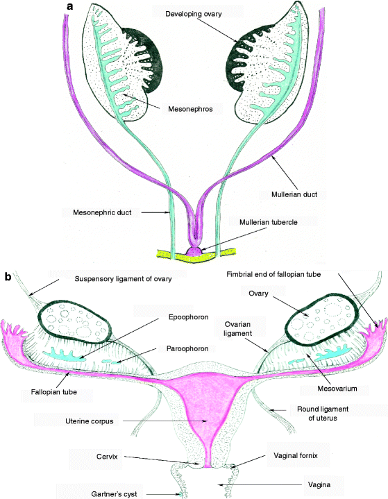
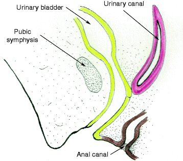
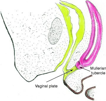
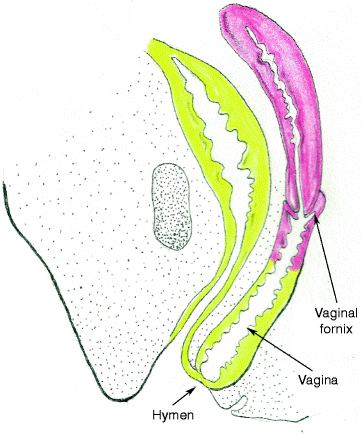
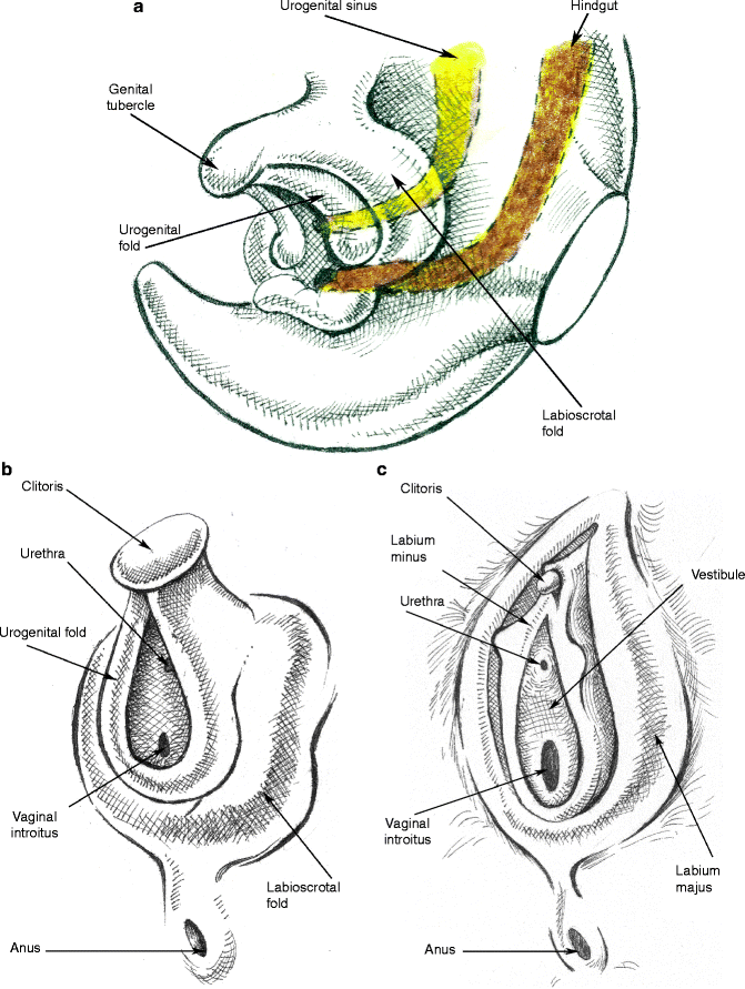
Pathology of the Vulva and Vagina introduces the topic with a stylishly illustrated description of the embryology and development which is fundamental to understanding the pathogenesis and symptomatology. Subsequent chapters cover infections and non-infectious dermatoses, specifying those that can predispose to cancer. The precancerous conditions of vulval intraepithelial neoplasia, melanocytic proliferations and extra-mammary Pagets disease are integrated respectively with accounts of human papilloma virus, malignant melanoma and recent awareness of ano-genital mammary-like glands. Advances in the recognition of potentially confusing benign conditions, prognosis and staging update the pathology of squamous and adenocarcinoma in these organs. The difficulties of sentinel node biopsy are explored and a comprehensive chapter clearly highlights the difficult differential diagnosis of mesenchymal lesions.

As most histopathology departments receive many gynecological specimens, Pathology of the Vulva and Vagina has been written to be useful diagnostically to general as well as specialist gynecological histopathologists and pathologists in training. Gynecologists, oncologists, dermatologists, genitourinary physicians and cancer nurse specialists will find expert insights here that will help in treatment and counselling of their patients.

Laurence Brown (editor): author's other books


Who wrote Pathology of the Vulva and Vagina? Find out the surname, the name of the author of the book and a list of all author's works by series.

Pathology of the Vulva and Vagina — read online for free the complete book (whole text) full work

Below is the text of the book, divided by pages. System saving the place of the last page read, allows you to conveniently read the book "Pathology of the Vulva and Vagina" online for free, without having to search again every time where you left off. Put a bookmark, and you can go to the page where you finished reading at any time.

Light

Font size:

Reset

Interval:

Bookmark:

Make
Laurence Brown (ed.) Essentials of Diagnostic Gynecological Pathology Pathology of the Vulva and Vagina 2013 10.1007/978-0-85729-757-0_1 Springer-Verlag London 2013
1. Development and Anatomy: Disorders of Development
Naveena Singh 1
(1)
Department of Cellular Pathology, Barts Health NHS Trust, 2nd Floor, 80 Newark Street, London, E1 2ES, UK
Naveena Singh
Email:
Abstract
An overview of the complex embryological development of the lower female genital tract, some of which remains incompletely understood, is useful for the understanding of many neoplastic and nonneoplastic vulvovaginal lesions. The Fallopian tubes, uterus, and cervix develop from the paramesonephric (Mullerian) ducts. The vagina has a dual origin with the upper portion, including the vaginal fornices, arising from the paramesonephric ducts and the lower portion from the urogenital sinus. The development of the mesoderm surrounding the female genital tract in humans is incompletely understood. Experimental studies demonstrate that complex stromal epithelial interactions are crucial for site-specific epithelial differentiation, especially for development of glandular epithelia. Development of external genitalia occurs under the influence of sex hormones through proliferation of the mesoderm and ectoderm lateral and ventral to the cloaca. The area bounded by the vaginal orifice and the urogenital sinus enlarges to form the vestibule and is of endodermal origin. This is morphologically and functionally distinct from the rest of vulval tissues which are mesodermally and ectodermally derived. This difference in origin is reflected in differences in responses to sex hormones and other stimuli. A variety of abnormalities in development can therefore occur as a result of structural and hormonal disturbances, including external influences such as in utero exposure to diethylstilbestrol.
Development of the Vulva and Vagina
An overview of the embryological development of the lower female genital tract is useful for the understanding of many neoplastic and nonneoplastic lesions that occur at this site. Although there have been many advances in the study of the development of the human embryo, the development of parts of urogenital system remains incompletely understood [].
The Fallopian tubes, uterus, and cervix develop from the paramesonephric (Mullerian) ducts. The vagina develops by contributions from the Mullerian ducts as well as the urogenital sinus. The paired Mullerian ducts originate in the fifth week as longitudinal grooves in the urogenital ridge, lying lateral to the mesonephric (Wolffian) ducts. The ducts grow caudally by cellular proliferation as solid tubes which canalize as they elongate. Caudally, these cross to the midline in front of (ventral to) the mesonephric ducts. The Mullerian ducts approach, fuse with each other, forming the uterovaginal canal, and join with the urogenital sinus in the seventh week (Fig. ].
Fig 11 a Mullerian and paramesonephric duct development b The female - photo 1
Fig. 1.1
( a ) Mullerian and paramesonephric duct development. ( b ) The female genital tract fully developed with remnants of the mesonephric ducts in blue
Around day 66, there is an upward migration of squamous epithelial cells from the urogenital sinus. This structure, initially a solid cylinder of cells which later canalizes, is known as the vaginal plate and is unique to the human embryo [).
Fig 12 Sagittal section through the developing uterovaginal canal at 9 weeks - photo 2
Fig. 1.2
Sagittal section through the developing uterovaginal canal at 9 weeks showing the Mullerian tubercle of paramesonephric and vaginal plate of urogenital sinus origin
Fig 13 The vagina at the end of the third month Fig 14 The fully - photo 3
Fig. 1.3
The vagina at the end of the third month
Fig 14 The fully canalized vagina in the newborn female More proximally - photo 4
Fig. 1.4
The fully canalized vagina in the newborn female
More proximally endocervical mucinous glands appear between the 13th and 15th weeks together with differentiation of endometrial and serous epithelia in the upper genital tract. The development of the mesoderm surrounding the female genital tract in humans is incompletely understood, but experimental studies demonstrate that complex stromal epithelial interactions are crucial for site-specific epithelial differentiation []. The outer layer forms the muscle wall of the Fallopian tube, the myometrium, the outer cervical stroma, and the muscle wall of the vagina.
Development of external genitalia occurs under the influence of sex hormones [). The ventral portions of the labioscrotal folds fuse medially to produce the mons pubis. The labioscrotal folds fuse posteriorly within the urogenital membrane anterior to the anus.
Fig 15 a Development of the external genitalia begins with the appearance - photo 5
Fig. 1.5
( a ) Development of the external genitalia begins with the appearance of the genital tubercle, labioscrotal fold, and urogenital fold; the cloaca has divided into the urogenital sinus and the hindgut. ( b ) The labioscrotal and urogenital folds remain separate in the female, enclosing the vestibule which receives the openings of the urethra and vagina (in the male, these pairs of folds fuse to form the scrotum). ( c ) The genital tubercle, urogenital folds, and labioscrotal folds give rise to the clitoris, labia minora, and labia majora, respectively
The area bounded by the vaginal orifice and the urogenital sinus enlarges to form the vestibule (see below), which is therefore largely of endodermal origin apart from a variable area anterior to the urethra. This is morphologically and functionally distinct from the rest of vulval tissues which are mesodermally and ectodermally derived, reflected in differences in responses to sex hormones and other stimuli. Typically, irritation in endodermally derived areas is perceived as burning in contrast to the itching felt in ectodermal epithelia.
Anatomy and Histology of the Vagina
The vagina is an anteroposteriorly collapsed hollow fibromuscular tube about 9 cm in length in the adult female. This opens into the vulva inferiorly. Superiorly, the vagina communicates with the cervix. The axis of the vagina lies in an oblique line with the top of the vagina lying superior and posterior to the introitus. The axis of the normally anteverted uterus and cervix forms an obtuse angle with that of the vagina, such that the external os of the cervix faces the posterior vaginal wall. The cervix bulges into the upper vagina and the blind folds of vaginal mucosa that surround the cervix are known as the fornices, being deepest posteriorly.
The inner surface of vagina is covered by a dull lining that bears many horizontal folds or rugae to enable stretching. There are normally no glands in the vagina and the secretions that are seen as part of the arousal response appear to be derived from transudate resulting from increased blood flow in mural vessels, with contributions from Bartholins, sebaceous, sweat, Skenes, and endocervical glands []. The vaginal fluid has a slightly acidic pH which varies with the phase of the menstrual cycle and during the arousal response when it has a higher pH and is rich in enzymes and immunoglobulins. The vaginal wall becomes more soft and relaxed during pregnancy and parturition.
Next page
Light

Font size:

Reset

Interval:

Bookmark:

Make

Similar books «Pathology of the Vulva and Vagina»

Look at similar books to Pathology of the Vulva and Vagina. We have selected literature similar in name and meaning in the hope of providing readers with more options to find new, interesting, not yet read works.


Reviews about «Pathology of the Vulva and Vagina»

Discussion, reviews of the book Pathology of the Vulva and Vagina and just readers' own opinions. Leave your comments, write what you think about the work, its meaning or the main characters. Specify what exactly you liked and what you didn't like, and why you think so.