Lichen Planus (LP)
Clinical : Commonly remembered as the five Ps purple, polygonal, planar, and pruritic papules (and/or plaques) with a fine overlying scale containing thin white lines (known as Wickhams striae ). LP commonly manifests as grouped papules on the distal extremities and/or trunk, also with frequent involvement of oral mucosa.
Hypertrophic Lichen Planus Typical inflammatory pattern with marked acanthosis of the epidermis which may mimic prurigo nodule or keratoacanthoma.
Lichen Planopilaris Arises on the scalp and causes a scarring alopecia (discussed more in detail in the alopecia chapter).
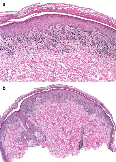
Histopathology : Compact orthokeratotic hyperkeratosis except for oral lesions which may have parakeratosis (Fig. ). Classically shows wedge-shaped hypergranulosis and irregular acanthosis of the epidermis ( saw-toothed rete ridges ). Underlying the epidermis is a band-like lymphocytic infiltrate which extends into the superficial epidermis causing vacuolar interface change . Colloid or civatte bodies can be found in the areas of liquefaction degeneration and sometimes in the superficial dermis. In more advanced lesions, the epidermis may become somewhat atrophic, and there may be a significant degree of pigment incontinence with dermal melnophages.
Fig. 1.1
( a ) There is a lichenoid lymphocytic infiltrate with saw-toothed rete ridges. There is also confluent orthokeratosis (hematoxylin and eosin, 10). ( b ) Lichen planus is the prototype for lichenoid infiltrates. There is a superficial band-like lymphocytic infiltrate which obscures the dermoepidermal junction (hematoxylin and eosin, 4). ( c ) Pigment incontinence and melanophages are frequently seen. (hematoxylin and eosin, 20)
Ancillary Studies : Direct immunofluorescence studies may show positive IgM, complement, and fibrin in the colloid bodies .
Differential Diagnosis : benign lichenoid keratosis, lichenoid actinic keratosis, halo nevus, lichenoid drug eruption, lupus erythematosus, erythema multiforme.
Benign Lichenoid Keratosis (Lichen Planus-Like Keratosis)
Clinical : Usually a solitary erythematous or violaceous papule which may arise on the arms or trunk. Patients may complain of pain, burning, or pruritis.
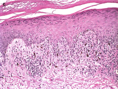
Histopathology : Nearly identical to lichen planus, except it is a solitary lesion (Fig. ). BLK represents a lichenoid immune reaction to a previously existing lesion (like seborrheic keratosis or solar lentigo). If there is keratinocytic atypia, then a dysplastic lesion (such as lichenoid AK) must be excluded. A careful search for a melanocytic proliferation should be negative.
Fig. 1.2
( a ) A lichenoid keratosis has a lichenoid lymphocytic infiltrate with saw-toothed epidermis at low power (hematoxylin and eosin, 4). ( b ) Civatte bodies, confluent orthokeratosis, a lichenoid lymphocytic infiltrate, and an irregular dermoepidermal junction can be seen. (hematoxylin and eosin, 20)
Differential Diagnosis : Lichen planus, lichenoid actinic keratosis, halo nevus, lichenoid drug eruption, lupus erythematosus, erythema multiforme.
Lichen Striatus
Clinical : Usually a childhood disease which manifests as a linear plaque with overlying scale. There may be hypopigmentation or hyperpigmentation. Typically resolves after several months.
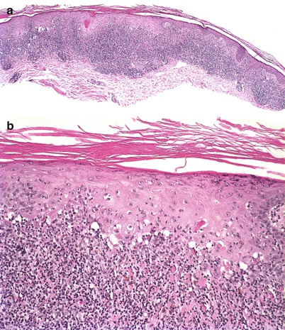
Histopathology : The epidermis is acanthotic or psoriasiform with overlying hyperkeratosis with or without focal parakeratosis (Fig. ). Spongiosis is a frequent finding. As in other lichenoid eruptions, there is vacuolar interface degeneration with dyskeratotic keratinocytes . The lymphocytic infiltrate is more perivascular, perifollicular, and, characteristically syringocentric (around the sweat ducts).
Fig. 1.3
( a ) The inflammation in lichen striatus also involves the underlying sweat glands in addition to the overlying lichenoid infiltrate (hematoxylin and eosin, 10). ( b ) There is a lichenoid lymphocytic infiltrate which also extends down to the sweat glands (hematoxylin and eosin, 4). ( c ) There is a lichenoid lymphocytic infiltrate superficially (hematoxylin and eosin, 10). ( d ) This is a close up view of the lymphocytic infiltrate within the sweat glands. (hematoxylin and eosin, 20)
Differential Diagnosis : Lichen planus, benign lichenoid keratosis, lichenoid actinic keratosis, halo nevus.
Lichen Nitidus
Clinical : Typically a childhood eruption of innumerable 12 mm skin-colored papules on the trunk and/or penis. Resolves after several months.
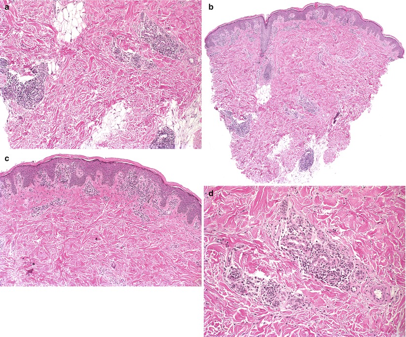
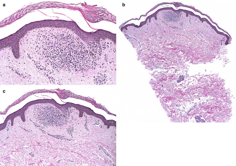
Histopathology : Dense lymphohistiocytic infiltrate in the superficial dermis which is localized or patchy (Fig. ). The overlying epidermis is usually atrophic with vacuolar interface change and lymphocytic infiltration. The epidermal retia are elongated around the lymphohystiocytic infiltrate forming a collarette the so-called ball in claw lesion. Multinucleated giant cells may also be present.
Fig. 1.4
( a ) The epidermal retia are beginning to extend around the granuloma. This is the beginning of the ball and clutch that is typical of lichen nitidus (hematoxylin and eosin, 10). ( b ) There is a small granuloma located immediately beneath the epidermis (hematoxylin and eosin, 4). ( c ) The epidermal retia are beginning to extend around the granuloma. This is the beginning of the ball and clutch that is typical of lichen nitidus. (hematoxylin and eosin, 10)
Differential Diagnosis : Lichen planus, benign lichenoid keratosis, lichenoid actinic keratosis, halo nevus.

