Enzo Berardesca (editor) - Non Invasive Diagnostic Techniques in Clinical Dermatology
Here you can read online Enzo Berardesca (editor) - Non Invasive Diagnostic Techniques in Clinical Dermatology full text of the book (entire story) in english for free. Download pdf and epub, get meaning, cover and reviews about this ebook. year: 2013, publisher: Springer, genre: Art. Description of the work, (preface) as well as reviews are available. Best literature library LitArk.com created for fans of good reading and offers a wide selection of genres:
Romance novel
Science fiction
Adventure
Detective
Science
History
Home and family
Prose
Art
Politics
Computer
Non-fiction
Religion
Business
Children
Humor
Choose a favorite category and find really read worthwhile books. Enjoy immersion in the world of imagination, feel the emotions of the characters or learn something new for yourself, make an fascinating discovery.

- Book:Non Invasive Diagnostic Techniques in Clinical Dermatology
- Author:
- Publisher:Springer
- Genre:
- Year:2013
- Rating:4 / 5
- Favourites:Add to favourites
- Your mark:
Non Invasive Diagnostic Techniques in Clinical Dermatology: summary, description and annotation
We offer to read an annotation, description, summary or preface (depends on what the author of the book "Non Invasive Diagnostic Techniques in Clinical Dermatology" wrote himself). If you haven't found the necessary information about the book — write in the comments, we will try to find it.
This book is a comprehensive but compact guide to the latest technical and technological developments in the growing field of non invasive diagnosis in clinical dermatology. Information is provided on the practical and technical characteristics of a wide range of equipment and methods for in vivo measurements that aid in the investigation of skin function, the evaluation of topically applied products and the monitoring of skin disease. Individual sections are devoted to imaging techniques, skin analysis, superficial skin analysis, skin mechanics, water and stratum corneum hydration and erythema and blood flow. All of the authors are experts in the field, with detailed knowledge of the techniques they describe. Non Invasive Diagnostic Techniques in Clinical Dermatology will be of value for all dermatologists, whether they are engaged in delivering patient care or in research programs, for cosmetic scientists and for biologists involved in skin research and product assessment.
Enzo Berardesca (editor): author's other books
Who wrote Non Invasive Diagnostic Techniques in Clinical Dermatology? Find out the surname, the name of the author of the book and a list of all author's works by series.

Non Invasive Diagnostic Techniques in Clinical Dermatology — read online for free the complete book (whole text) full work
Below is the text of the book, divided by pages. System saving the place of the last page read, allows you to conveniently read the book "Non Invasive Diagnostic Techniques in Clinical Dermatology" online for free, without having to search again every time where you left off. Put a bookmark, and you can go to the page where you finished reading at any time.
Font size:
Interval:
Bookmark:
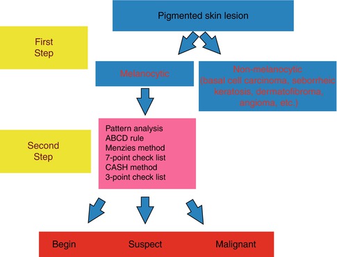
Imaging Techniques
Pigmented skin lesions |
Melanocytic (nevi, melanoma) |
Non-melanocytic (solar lentigo, seborrheic keratosis, dermatofibroma, basal cell carcinoma) |
Nonpigmented skin lesions |
Sebaceous hyperplasia |
Pyogenic granuloma |
Clear cell acanthoma |
Xanthomatous neoplasms |
Mastocytosis |
Sarcoidosis |
Median raphe cysts |
Eccrine poroma |
Keratoacanthoma |
Actinic porokeratosis |
Nonpigmented facial actinic keratosis |
Bowens disease |
Invasive squamous cell carcinoma |
Kaposis sarcoma |
Ectoparasitoses |
Scabies |
Head and pubic lice |
Tungiasis |
Cutaneous leishmaniasis |
Furuncular myiasis |
Demodicosis |
Cutaneous / mucosal infections |
Molluscum contagiosum |
Cutaneous warts |
Genital warts |
Tinea nigra |
Lupus vulgaris |
Inflammatory disorders |
Psoriasis |
Lichen planus |
Urticaria and urticarial vasculitis |
Rosacea |
Pityriasis lichenoides et varioliformis acuta |
Scalp disorders (see ) |
Hair loss |
Parasitoses |
Psoriasis |
Hair shaft disorders |
Nail disorders |
Psoriasis |
Onychomycosis |
Onychomatricoma |
Glomus tumor |
Vascular disorders |
Port-wine stains |
Infantile hemangioma |
Pigmented purpuric dermatoses (PPD) |
VD features | Description | Histopathologic correlation | |
|---|---|---|---|
Pigment network | Grid of brownish lines over a tan background. | The lines correspond to melanin pigment contained in keratinocytes or in melanocytes outlining the pattern of the epidermal rete ridges, while the tan areas among them correspond to the dermal papillae tips | 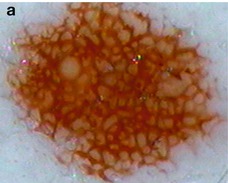 |
It can be typical (regular, thin, narrow) or atypical (irregular, thick, wide) in benign and malignant melanocytic lesions, respectively | |||
Diffuse pigmentation | According to the localization of the melanin pigment within the skin, different colors can be seen. | Melanin pigment localized within the epidermal stratum corneum appears black; in the lower epidermal layers light to dark brown; in the papillary dermis gray; pigmentation of the reticular dermis is steel blue | 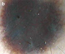 |
Even and uneven pigmentation is usually found in benign and malignant lesions | |||
Hypopigmentation | Diffuse or localized areas of decreased pigmentation, commonly observed in benign melanocytic lesions | Decreased melanin pigment | 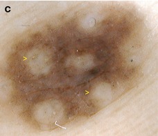 |
Font size:
Interval:
Bookmark:
Similar books «Non Invasive Diagnostic Techniques in Clinical Dermatology»
Look at similar books to Non Invasive Diagnostic Techniques in Clinical Dermatology. We have selected literature similar in name and meaning in the hope of providing readers with more options to find new, interesting, not yet read works.
Discussion, reviews of the book Non Invasive Diagnostic Techniques in Clinical Dermatology and just readers' own opinions. Leave your comments, write what you think about the work, its meaning or the main characters. Specify what exactly you liked and what you didn't like, and why you think so.