• Complain

Plaza - Pathology of Pigmented Skin Lesions

Here you can read online Plaza - Pathology of Pigmented Skin Lesions full text of the book (entire story) in english for free. Download pdf and epub, get meaning, cover and reviews about this ebook. City: Berlin;Heidelberg, year: 2018, publisher: Springer-Verlag Berlin An, genre: Home and family. Description of the work, (preface) as well as reviews are available. Best literature library LitArk.com created for fans of good reading and offers a wide selection of genres:

Romance novel Science fiction Adventure Detective Science History Home and family Prose Art Politics Computer Non-fiction Religion Business Children Humor

Choose a favorite category and find really read worthwhile books. Enjoy immersion in the world of imagination, feel the emotions of the characters or learn something new for yourself, make an fascinating discovery.

Plaza Pathology of Pigmented Skin Lesions

Pathology of Pigmented Skin Lesions: summary, description and annotation

We offer to read an annotation, description, summary or preface (depends on what the author of the book "Pathology of Pigmented Skin Lesions" wrote himself). If you haven't found the necessary information about the book — write in the comments, we will try to find it.

Plaza: author's other books


Who wrote Pathology of Pigmented Skin Lesions? Find out the surname, the name of the author of the book and a list of all author's works by series.

Pathology of Pigmented Skin Lesions — read online for free the complete book (whole text) full work

Below is the text of the book, divided by pages. System saving the place of the last page read, allows you to conveniently read the book "Pathology of Pigmented Skin Lesions" online for free, without having to search again every time where you left off. Put a bookmark, and you can go to the page where you finished reading at any time.

Light

Font size:

Reset

Interval:

Bookmark:

Make
Springer-Verlag Berlin Heidelberg 2017
Jose A. Plaza and Victor G. Prieto Pathology of Pigmented Skin Lesions 10.1007/978-3-662-52721-4_1
1. Lentigines
Jose A. Plaza 1 and Victor G. Prieto 2
(1)
Division of Dermatopathology, Miraca Life Sciences, Dallas, TX, USA
(2)
MD Anderson Cancer Center, University of Texas, Houston, TX, USA
Ephelides
Clinical Features
Ephelides (freckles) are common, uniformly pigmented, multiple macules (15 mm in size) mainly limited to body regions above the waist. These macules are more numerous on sun-exposed areas (face, shoulders, and upper back) and usually fade and become smaller in the winter season. Ephelides appear early in childhood and partly disappear with age and are closely related to pigmentary host characteristics such as fair skin and/or red hair. Only rarely ephelides are seen in individuals with dark skin. Ephelides may manifest an autosomal dominant pattern of inheritance (appearing in sequential generations). High levels of freckling may indicate a raised susceptibility to the development of melanoma. In contrast to solar lentigines, ephelides are not strongly associated with age [].
Histopathology
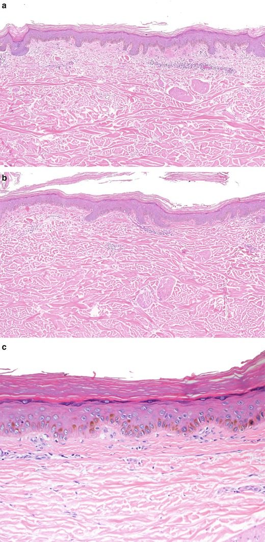
The epidermis appears normal in structure. The basal cells in the affected areas are more heavily pigmented with melanin than those in the surrounding skin, and there is usually sharp delimitation of the abnormal from the normal areas. There are normal and sometimes decreased numbers of melanocytes within epidermis; however, the melanosomes in those cells are larger than those in the surrounding skin and can sometimes be seen with the microscope as dark, large, intracytoplasmic granules (macromelanosomes). The adnexal structures are not involved. By electron microscopy studies, the melanocytes contain enlarged spherical granular melanosomes as opposed to the striated ellipsoid forms seen in normal skin [ac).
Fig 11 Ephelides Note the pigmented basal keratinocytes and the decreased - photo 1
Fig. 1.1
Ephelides. Note the pigmented basal keratinocytes and the decreased number of melanocytes within epidermis ( a ). Higher magnification showing melanocytes with rare melanosomes ( b ). Observe the lack of melanocytes ( c )
Differential Diagnosis
The clinical differential diagnosis of ephelides includes lentigo simplex and caf au lait spot. The diagnosis of caf au lait spot will rely on the identification of giant melanosomes and mild increased number of melanocytes. Lentigo simplex (as explained in detail below) will show elongated rete ridges with hyperpigmentation of basal keratinocytes (some authors accept also mild increased in numbers of melanocytes) (Fig. ac).
Lentigo Simplex
Clinical Features
Lentigo simplex is a very common, benign, pigmented lesion that can be found anywhere on the body surface and is preferentially observed in young people, although it may occur at any age. They usually appear early in life and are typically not associated with sun exposure. Clinically, lesions present as non-palpable, relatively symmetric, uniform, homogeneous, light-brown to black macules, on the trunk, extremities, genitals, and mucosa surfaces, usually measuring less than 5 mm in size (in certain anatomic sites such as the palms, soles, genitalia, and mucosal membranes, they can be larger). It is rare for a lentigo simplex to be asymmetrical or to have irregular borders. Lentigo simplex may occur as single or multiple lesions. It has been proposed that lentigo simplex may evolve into a lentiginous/junctional melanocytic nevus when melanocytes start proliferating and aggregating to form small nests in the junctional zone. On the other hand, a recent study has shown that absence of BRAF, FGFR3, and PIK3CA mutations can clearly differentiate lentigo simplex from melanocytic nevi and solar lentigo. These results furthermore indicate that lentigo simplex has a distinct yet unknown genetic basis, which does not necessarily exclude the proposed lentigo-nevus sequence [].
In some instances, multiple lentigines are associated with rare genetic disorders. These include LEOPARD syndrome (lentigines, EKG changes, ocular hypertelorism, pulmonary stenosis, abnormal genitalia, growth retardation, and deafness), Carney complex (lentigines, atrial myxoma, mucocutaneous myxoma, and nevi), Peutz-Jeghers syndrome (oral and perioral lentigines, multiple gastrointestinal polyps, and visceral tumors), xeroderma pigmentosum (lentigines on sun-exposed skin and multiple skin cancers), and Cronkhite-Canada syndrome (buccal, facial, and palmoplantar lentigines, alopecia, nail dystrophy, and intestinal polyps).
Histopathology
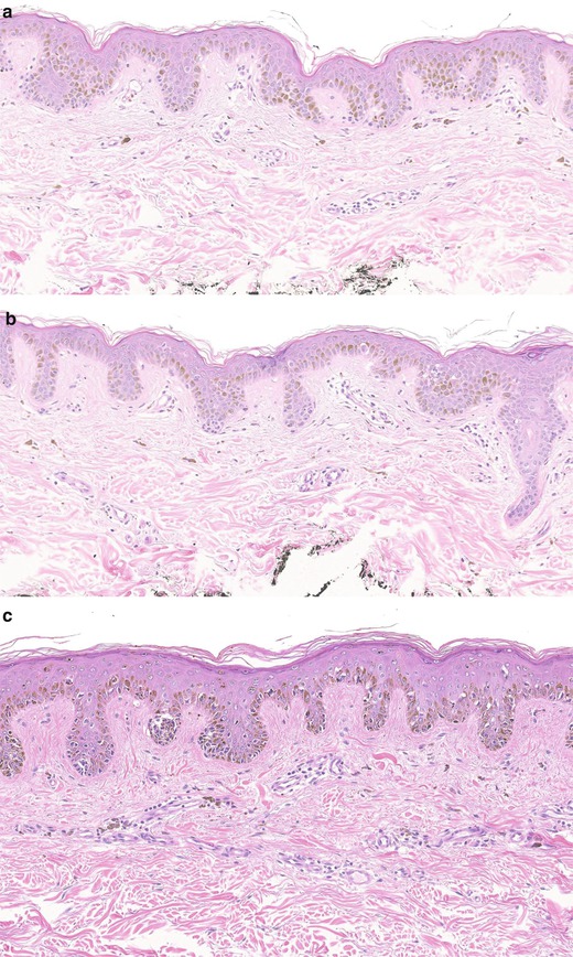
Lentigo simplex shows mild elongation of the epidermal rete ridges with variable basal cell hyperpigmentation. The elongated rete ridges are either thin or club shaped. Some authors accept that there may be an increased number of single melanocytes in the basal layer devoid of cytologic atypia. In some cases there are giant melanosomes as in lentigines. Papillary dermis is often fibrotic; however, in contrast with dysplastic rarely is there concentric and lamellar fibroplasia. Also in the papillary dermis, there may be a subtle lymphohistiocytic infiltrate with scattered melanophages. While the majority of lentigo simplex are stable, some may develop junctional nests and melanocytes may descent into papillary dermis. Thus, lentigos and junctional nevi may coexist and can be designated as lentiginous junctional nevus (jentigo) [ac).
Fig 12 Lentigo simplex The lesion shows elongation of rete ridges and - photo 2
Fig. 1.2
Lentigo simplex. The lesion shows elongation of rete ridges and pigmented keratinocytes with absence of solar damage ( a ). The pigment varies within epidermis and can be prominent in some cases ( b ). An early transition into an early junctional nevus (jentigo) ( c )
Differential Diagnosis
Lentigo simplex is often biopsied to rule out melanoma in situ, especially if the lesion is located in the head and neck of an older individual. In most cases, this differential diagnosis is straightforward. However, in some instances, lentigo simplex may display isolated melanocytes at and above the basal layer. This represents a rare occurrence, and its distinction with melanoma in situ can be problematic if the specimen is small and the complete architecture of the lesion cannot be evaluated. Lentigo simplex located in special sites (such as umbilical, genital, and axillary areas) can also show irregular distribution of the pigment. Close attention to the cytology of the cells is very important to make a distinction from melanoma, as these melanocytes do not differ from those seen in the basal layer of the epidermis, and cytologically they have small nuclei with compact chromatin and scant cytoplasm. Upwardly scattered melanocytes can also be seen in cases of lentigo simplex that have been traumatized. The presence of parakeratosis, spongiosis, extravasated red blood cells, dyskeratotic keratinocytes, and the melanin pigment above the suprabasal layer is a hint that a lesion has been traumatized. The presence of melanocytes with vesicular nuclei and abundant cytoplasm along with signs of solar damage, pagetoid spread of melanocytes, irregular distribution of melanin pigment in epidermis and dermis, and a dense lichenoid inflammatory infiltrate in superficial dermis (especially underneath the area of the lesion) raises the possibility of melanoma in situ. A diagnostic hallmark of melanoma in situ (especially lentigo maligna) is the extension of neoplastic melanocytes down the adnexal epithelial structures , a phenomenon not seen in lentigo simplex (Fig. ac).
Next page
Light

Font size:

Reset

Interval:

Bookmark:

Make

Similar books «Pathology of Pigmented Skin Lesions»

Look at similar books to Pathology of Pigmented Skin Lesions. We have selected literature similar in name and meaning in the hope of providing readers with more options to find new, interesting, not yet read works.


Reviews about «Pathology of Pigmented Skin Lesions»

Discussion, reviews of the book Pathology of Pigmented Skin Lesions and just readers' own opinions. Leave your comments, write what you think about the work, its meaning or the main characters. Specify what exactly you liked and what you didn't like, and why you think so.