Jose A. Plaza - Pathology of Pigmented Skin Lesions
Here you can read online Jose A. Plaza - Pathology of Pigmented Skin Lesions full text of the book (entire story) in english for free. Download pdf and epub, get meaning, cover and reviews about this ebook. year: 2017, publisher: Springer Berlin Heidelberg, genre: Home and family. Description of the work, (preface) as well as reviews are available. Best literature library LitArk.com created for fans of good reading and offers a wide selection of genres:
Romance novel
Science fiction
Adventure
Detective
Science
History
Home and family
Prose
Art
Politics
Computer
Non-fiction
Religion
Business
Children
Humor
Choose a favorite category and find really read worthwhile books. Enjoy immersion in the world of imagination, feel the emotions of the characters or learn something new for yourself, make an fascinating discovery.

- Book:Pathology of Pigmented Skin Lesions
- Author:
- Publisher:Springer Berlin Heidelberg
- Genre:
- Year:2017
- Rating:3 / 5
- Favourites:Add to favourites
- Your mark:
Pathology of Pigmented Skin Lesions: summary, description and annotation
We offer to read an annotation, description, summary or preface (depends on what the author of the book "Pathology of Pigmented Skin Lesions" wrote himself). If you haven't found the necessary information about the book — write in the comments, we will try to find it.
Jose A. Plaza: author's other books
Who wrote Pathology of Pigmented Skin Lesions? Find out the surname, the name of the author of the book and a list of all author's works by series.


Pathology of Pigmented Skin Lesions — read online for free the complete book (whole text) full work
Below is the text of the book, divided by pages. System saving the place of the last page read, allows you to conveniently read the book "Pathology of Pigmented Skin Lesions" online for free, without having to search again every time where you left off. Put a bookmark, and you can go to the page where you finished reading at any time.
Font size:
Interval:
Bookmark:


This Springer imprint is published by Springer Nature
The registered company is Springer-Verlag GmbH Germany
The registered company address is: Heidelberger Platz 3, 14197 Berlin, Germany
The histomorphologic study of pigmented lesions of the skin constitutes the majority of our daily dermatopathology practice. In this book, we have compiled our experience in regard to pigmented lesions of the skin and have illustrated not only stereotypical examples of pigmented lesions but also unusual variants and a wide spectrum of variations that can be observed in such lesions. Our goal in this book is to illustrate thoroughly the most difficult topics in melanocytic tumors and to describe our view on how to diagnose these lesions. The opinions stated in this book represent our personal views on the topics. As with other topics in the field of pathology, the reader should consider the information provided and assimilate it to her/his own experience.
The histological diagnosis of melanocytic lesions is one of the most difficult areas in pathology, given the subjectivity and histologic variations that some of such entities may depict. In the last decade, there have been major advances in terms of diagnosis and prognosis of such lesions. It is well known that a number of these lesions cannot be precisely and reproducibly classified as either entirely benign or malignant just by using conventional histologic and immunohistochemical techniques. New understandings of molecular pathogenesis of melanocytic proliferations have revealed genetic differences between nevi and melanoma that can be used as targets for developing molecular diagnostic tests. FISH has emerged as a preferred molecular technique to interrogate chromosomal abnormalities, with proven utility as a diagnostic adjunct in lymphoid lesions and solid tumors and that has been recently validated for the diagnosis of melanocytic lesions. In this book, in addition to special mention to immunohistochemistry, we cover also the utility of adjunct molecular studies applied to the diagnosis of certain melanocytic lesions that are exceedingly difficult to diagnose on pure histomorphologic grounds.
We are in debt with our teachers, colleagues, and students who have guided and challenged us over the years. We also are thankful to many pathologists who have shared their challenging cases with us on consultation, and we have learned from them. The major sources of material used in book are from the dermatopathology divisions of Medical College of Wisconsin, University of Texas MD Anderson Center, and Miraca Life Sciences. And last, but not least, we are much in debt with our families who have supported us during all these years.
Ephelides (freckles) are common, uniformly pigmented, multiple macules (15 mm in size) mainly limited to body regions above the waist. These macules are more numerous on sun-exposed areas (face, shoulders, and upper back) and usually fade and become smaller in the winter season. Ephelides appear early in childhood and partly disappear with age and are closely related to pigmentary host characteristics such as fair skin and/or red hair. Only rarely ephelides are seen in individuals with dark skin. Ephelides may manifest an autosomal dominant pattern of inheritance (appearing in sequential generations). High levels of freckling may indicate a raised susceptibility to the development of melanoma. In contrast to solar lentigines, ephelides are not strongly associated with age [].
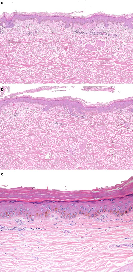
Ephelides. Note the pigmented basal keratinocytes and the decreased number of melanocytes within epidermis (a). Higher magnification showing melanocytes with rare melanosomes (b). Observe the lack of melanocytes (c)
The clinical differential diagnosis of ephelides includes lentigo simplex and caf au lait spot. The diagnosis of caf au lait spot will rely on the identification of giant melanosomes and mild increased number of melanocytes. Lentigo simplex (as explained in detail below) will show elongated rete ridges with hyperpigmentation of basal keratinocytes (some authors accept also mild increased in numbers of melanocytes) (Fig. ac).
Font size:
Interval:
Bookmark:
Similar books «Pathology of Pigmented Skin Lesions»
Look at similar books to Pathology of Pigmented Skin Lesions. We have selected literature similar in name and meaning in the hope of providing readers with more options to find new, interesting, not yet read works.
Discussion, reviews of the book Pathology of Pigmented Skin Lesions and just readers' own opinions. Leave your comments, write what you think about the work, its meaning or the main characters. Specify what exactly you liked and what you didn't like, and why you think so.