1. Immunohistology and Molecular Studies of Epithelial Tumors
Basal Cell Carcinoma
Basal cell carcinoma (BCC)is the most common cutaneous neoplasm. In one population-based study in Rochester, Minnesota, the annual incidence for BCC was estimated at 146 cases per 100,000 persons [].
Histologically, cells of BCC resemble cells of the basal layer of the epidermis; however, the cell of origin for BCC remains unclear and controversial. The varying histologic subtypes may arise from different cellular compartments of the skin, particularly stem cells of the interfollicular epidermis and from the upper infundibulum [].
The nevoid basal cell carcinoma syndrome (NBCCS) is an autosomal disorder manifesting as hundreds to thousands of BCCs in a single patient in addition to odontogenic keratocysts, palmar and plantar pits, and increased incidence of other neoplasms including medulloblastomas and rhabdomyosarcomas [].
On histology, BCC shows nests of basaloid keratinocytes surrounded by stoma, often with peritumoral retraction and peripheral palisading of lesional cells. Mucin is often present. Mitotic and apoptotic cells are frequent. Different subtypes of BCC demonstrate different histologic patterns.
Superficial BCC shows connection to the epidermis with downward buds of basaloid cells, often with peritumoral retraction. Nodular BCC shows nests, often large, that extend into the papillary and reticular dermis. Peritumoral retraction and mucin pooling is frequent. Morpheaform BCC shows infiltrative strands of BCC within a thick, collagenous stroma, that can often extend into the deep dermis and subcutis. Micronodular BCC shows small round nests of BCC with an associated dense stroma that can extend deep into the dermis or subcutis. Basosquamous BCC or metatypical BCC shows a basaloid cell morphology but with areas of keratinization.
The diagnosis of BCC with an adequate sample is often straightforward without any immunohistochemical stain required. Different subtypes of BCC, however, on a superficial shave biopsy, may raise the possibility of other diagnoses that require alternative treatment modalities. In such circumstances, IHC can be applied, in addition to morphologic criteria, to favor one diagnosis over the other.
Infiltrative BCC, microcystic adnexal carcinoma (MAC) , and desmoplastic trichoepithelioma (DTE) can show morphologic similarities and immunohistochemistry stains are useful in this differential. BerEP4 shows positivity in BCC and DTE, whereas MAC is typically negative. BerEP4, however, can be negative in the center or surface of BCC and in areas with a more squamoid morphology [).
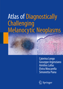
Fig. 1.1
( a ) This is an example of infiltrative BCC. In small or in limited tissue samples, lesions can show similarities with a desmoplastic trichoepithelioma. ( b ) Infiltrative BCC composed of small cords and fascicles of basaloid cells
BCC with squamous metaplasia versus SCC with basaloid features can be a difficult differential on histologic grounds. BCC shows strong and diffuse staining for both CK17 and CK14, in addition to Ber-EP4 and although SCC can show occasional positivity for each marker individually, it should not show diffuse positivity for all markers [].
Sebaceous carcinoma (SC) is an aggressive neoplasm that may histologically mimic other malignant tumors, especially BCC and SCC; therefore, its recognition is important for appropriate treatment. In the past, Oil Red O was used to identify intracytoplasmic lipids in sebaceous lesions in fresh frozen tissue; however, standard permanent tissue processing extracts the intracellular lipids and often nonspecifically highlights macrophages and non-lipid laden cells in the background stroma. Many studies have analyzed conventional markers such as EMA, Ber-EP4, androgen receptor, CK7, and P53 [).
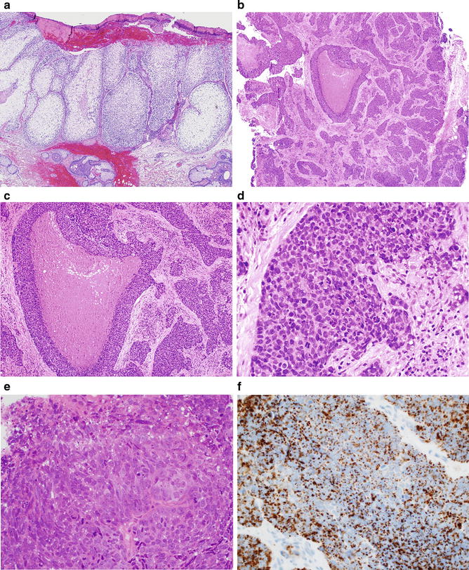
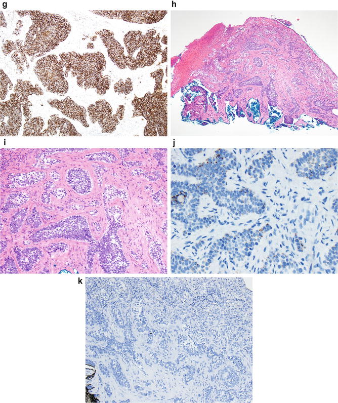
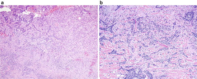
Fig. 1.2
( a ) This is an example of a low grade sebaceous carcinoma. These neoplasms are usually easily diagnosed on H&E and do not need IHC workup. ( b ) This a poorly differentiated sebaceous carcinoma. Note the basaloid features and the comedo necrosis. These neoplasms can be confused with other malignancies such as basal cell carcinoma and squamous cell carcinoma. ( c ) High power of a poorly differentiated sebaceous carcinoma. Note the lack of sebaceous differentiation requiring IHC to further classify the process. ( d ) Poorly differentiated sebaceous carcinoma. A diagnostic clue is the presence of a subtle vacuolar pattern in the cytoplasm of the neoplastic cells. ( e ) Another example of poorly differentiated sebaceous carcinoma. Note the subtle cytoplasmic vacuoles in the basaloid cells. ( f ) Cytoplasmic vacuolar pattern with adipophilin. ( g ) Sebaceous carcinoma. EMA is strongly positive. ( h ) This is a example of BCC with clear cell features. This neoplasm was localized in the eyelid of a 56-year-old man. The ddx of this neoplasm is with sebaceous carcinoma. ( i ) BCC with clear cell features. High power of the basaloid cells with clear cell differentiation. ( j ) BCC with clear cell features. Adipophilin stain is negative, but notice the focal granular staining that is classically seen in BCC. ( k ) BCC with clear cell features. EMA is negative
Another common problem in dermatopathology is the distinction between trichoepithelioma and BCC. Histomorphologic analysis has become in recent years more difficult because of the use of small shave biopsies, which for the most part deprive the histomorphologist of many architectural clues to the correct diagnosis. Trichoepitheliomas are hamartomatous tumors with follicular differentiation and a mesenchymal response equivalent to that seen in a normal hair follicle. Histopathologic features that favor a diagnosis of trichoepithelioma include a well circumscribed symmetrical lesion with symmetric and smooth margins, mesenchymal body differentiation usually in the form of germs and papillae, a cribriform pattern of neoplastic basaloid cells, clefts within the stroma and between the stroma and adjacent normal dermis, cornification of neoplastic epithelial cells that can be seen in association with foreign body granulomas, and a highly fibrotic stroma resembling that of perifollicular connective tissue. In contrast, histopathologic features that will favor a diagnosis of BCC, include an asymmetrical poorly circumscribed lesion, often with jagged borders, numerous necrotic neoplastic cells often en masse, frequent presence of melanin in germinative cells, frequent presence of acid mucosubstances within aggregations of epithelial cells and in the stroma, clefts between neoplastic cells and stroma, an edematous stroma, infiltrates of inflammatory cells, and frequent ulceration. Due to the major differences in management and prognosis of these two lesions, a correct diagnosis is desirable and expected. A number of studies have evaluated ancillary diagnostic tools, such as the immunohistochemical studies using CD34, CK15, CK14, Ber-Ep4, bcl-2, p53, etc., but have provided only limited support and diagnostic utility in the adjunctive histological separation of BCCs from trichoepitheliomas. Additionally, some studies have expressed the opinion that these two neoplasms may represent different points along the same neoplastic spectrum and are therefore difficult to differentiate on immunohistochemical techniques. We analyzed the expression of podoplanin (D240) in this setting. This antibody is a well-known lymphovascular marker that has been shown to react in the basal layer of the outer root sheath of hair follicles, peripheral germinative cells of sebaceous glands, and some cutaneous neoplasms. In our study, we found expression of podoplanin in 22.20% of BCCs and in 95% of trichoepitheliomas. Also, podoplanin expression in trichoepitheliomas was noted to be present not only in the basal layer of the tumor cells but also in the suprabasal layer of the tumor cells, as opposed to BCCs in which the positivity was primarily located in the basal layer of the tumor nests. The overall sensitivity and specificity of podoplanin immunoreactivity to separate primary BCCs from trichoepitheliomas was 95.5% and 77.8%, respectively, in this study []. The fact that a subset of BCCs can express podoplanin could embrace the premise that these neoplasms may have the potential for multilinear differentiation.