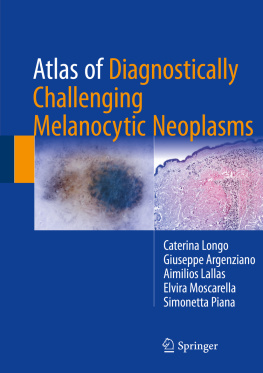
Caterina Longo - Atlas of Diagnostically Challenging Melanocytic Neoplasms
Here you can read online Caterina Longo - Atlas of Diagnostically Challenging Melanocytic Neoplasms full text of the book (entire story) in english for free. Download pdf and epub, get meaning, cover and reviews about this ebook. year: 2017, publisher: Springer, genre: Home and family. Description of the work, (preface) as well as reviews are available. Best literature library LitArk.com created for fans of good reading and offers a wide selection of genres:
Romance novel
Science fiction
Adventure
Detective
Science
History
Home and family
Prose
Art
Politics
Computer
Non-fiction
Religion
Business
Children
Humor
Choose a favorite category and find really read worthwhile books. Enjoy immersion in the world of imagination, feel the emotions of the characters or learn something new for yourself, make an fascinating discovery.

- Book:Atlas of Diagnostically Challenging Melanocytic Neoplasms
- Author:
- Publisher:Springer
- Genre:
- Year:2017
- Rating:3 / 5
- Favourites:Add to favourites
- Your mark:
Atlas of Diagnostically Challenging Melanocytic Neoplasms: summary, description and annotation
We offer to read an annotation, description, summary or preface (depends on what the author of the book "Atlas of Diagnostically Challenging Melanocytic Neoplasms" wrote himself). If you haven't found the necessary information about the book — write in the comments, we will try to find it.
Caterina Longo: author's other books
Who wrote Atlas of Diagnostically Challenging Melanocytic Neoplasms? Find out the surname, the name of the author of the book and a list of all author's works by series.


Atlas of Diagnostically Challenging Melanocytic Neoplasms — read online for free the complete book (whole text) full work
Below is the text of the book, divided by pages. System saving the place of the last page read, allows you to conveniently read the book "Atlas of Diagnostically Challenging Melanocytic Neoplasms" online for free, without having to search again every time where you left off. Put a bookmark, and you can go to the page where you finished reading at any time.
Font size:
Interval:
Bookmark:


This Springer imprint is published by Springer Nature
The registered company is Springer International Publishing AG
The registered company address is: Gewerbestrasse 11, 6330 Cham, Switzerland
I must definitely applaud the original idea of Caterina Longo of producing this atlas, which is a superb collection of cases outlining the diagnostic challenges we are facing every day with melanocytic tumors. For clinicians managing patients with melanoma, it is clear-cut that differentiating melanoma from benign melanocytic lesions can be very difficult at times. In the last 20 years, the practice changed dramatically because of the introduction of new tools for the preoperative diagnosis. Especially the introduction of dermoscopy and confocal microscopy allowed clinicians to improve their ability to recognize the many faces of melanocytic tumors with a quasi-histopathologic accuracy. However, there is a group of lesions that still are difficult to diagnose because of their equivocal clinical and/or histopathologic features. In these cases, only a careful clinic-pathologic correlation is the method to rich more closely the final diagnosis and, thus, the correct patient management. What I learned in the last 20 years is that nobody has 100% diagnostic accuracy, neither the clinician nor the pathologist. In difficult cases only an open-minded discussion among clinicians and pathologists is able to make the difference. Only by integrating all the possible information, the history, the clinical data, and the histopathologic features might we reach a more reasonable management of our patients. The aim of this book is indeed to illustrate this method, the clinic-pathologic correlation of difficult cases!
The sentence we are born and we will die without nevi summarizes one of the key components of the diagnosis of atypical lesions in the elderly. Epidemiologic data demonstrate that the nevus count and prevailing nevus patterns are strongly influenced by age. Notably, nevus count increases from childhood to midlife and decreases thereafter. In light of these findings, if evolving nevi in adolescence are an expected finding and therefore do not require further interventions, a melanocytic skin lesion showing signs of growth in the elderly should raise the index for malignancy. Furthermore, any flat acquired melanocytic lesion in this age should be considered with caution since the majority of lesions in the elderly are persistent intradermal nevi (congenital type). Firstly termed as atypical lentiginous junctional melanocytic proliferations, indeed they are regarded nowadays as melanomas.
Clinically, these atypical lentiginous junctional melanocytic proliferations of the elderly are commonly located on the upper back, shoulders, or extremities. They are solitary, often large (>8 mm), ill-defined macules with different shades of black, brown, and gray. Dermoscopically, these lesions are typified by a more or less atypical pigmented network, diffuse structureless brown pigmentation, and areas of regression. Studies employing digital dermoscopic follow-up suggest that these melanomas belong to a group of slow-growing tumors, which may grow in situ for several years. Their histologic diagnosis can be very difficult as they are cytologically bland and show scant epidermotropism and very little atypia. In the early phases, only a numerical increase of the melanocytes, scattered along the basal epidermis, can be noted. The alternation of single cells and irregular nests, with skip areas, usually within a sun-damaged skin, is an important histological clue for a diagnosis of early in situ melanoma.
What is a common finding in this age group is the presence of several benign non-melanocytic skin lesions such as seborrheic keratosis, angiomas, or solar lentigos. Thus, the clues to identify flat melanomas are the following: solitary flat pigmented lesions, large size, with network and regression on dermoscopy. Conversely, the presence of rough surface, comedo-like openings, fingerprinting, and red lacunae should be regarded as benign clues. However, the recognition of incipient melanomas should always be based on clinical data, patients phenotype, and history.
Font size:
Interval:
Bookmark:
Similar books «Atlas of Diagnostically Challenging Melanocytic Neoplasms»
Look at similar books to Atlas of Diagnostically Challenging Melanocytic Neoplasms. We have selected literature similar in name and meaning in the hope of providing readers with more options to find new, interesting, not yet read works.
Discussion, reviews of the book Atlas of Diagnostically Challenging Melanocytic Neoplasms and just readers' own opinions. Leave your comments, write what you think about the work, its meaning or the main characters. Specify what exactly you liked and what you didn't like, and why you think so.