• Complain

Luis Requena - Cutaneous Adnexal Neoplasms

Here you can read online Luis Requena - Cutaneous Adnexal Neoplasms full text of the book (entire story) in english for free. Download pdf and epub, get meaning, cover and reviews about this ebook. year: 2017, publisher: Springer, genre: Home and family. Description of the work, (preface) as well as reviews are available. Best literature library LitArk.com created for fans of good reading and offers a wide selection of genres:

Romance novel Science fiction Adventure Detective Science History Home and family Prose Art Politics Computer Non-fiction Religion Business Children Humor

Choose a favorite category and find really read worthwhile books. Enjoy immersion in the world of imagination, feel the emotions of the characters or learn something new for yourself, make an fascinating discovery.

Luis Requena Cutaneous Adnexal Neoplasms

Cutaneous Adnexal Neoplasms: summary, description and annotation

We offer to read an annotation, description, summary or preface (depends on what the author of the book "Cutaneous Adnexal Neoplasms" wrote himself). If you haven't found the necessary information about the book — write in the comments, we will try to find it.

This superbly illustrated book is the most comprehensive available guide to adnexal neoplasms of the skin. More than 70 entities are described in individual chapters that follow a uniform structure: historical review, clinical features, histopathology, histogenesis, immunohistochemistry, molecular anomalies, and treatment. Readers will find state of the art knowledge on all aspects, including the cytogenetic and chromosomal abnormalities associated with each neoplasm. Without exception, the illustrations are high-quality, full-color, original digital pictures. The histopathology images are taken from perfectly cut and stained sections and the immunohistochemistry illustrations are of an unrivalled quality among textbooks of dermatology and dermatopathology. A complete list of references from original description to the present day is also supplied for each neoplasm. Cutaneous adnexal neoplasms are a large and heterogeneous group of benign and malignant lesions. This book will assist the reader in early and correct recognition, which is essential for appropriate choice of treatment and prognostic assessment.

Luis Requena: author's other books


Who wrote Cutaneous Adnexal Neoplasms? Find out the surname, the name of the author of the book and a list of all author's works by series.

Cutaneous Adnexal Neoplasms — read online for free the complete book (whole text) full work

Below is the text of the book, divided by pages. System saving the place of the last page read, allows you to conveniently read the book "Cutaneous Adnexal Neoplasms" online for free, without having to search again every time where you left off. Put a bookmark, and you can go to the page where you finished reading at any time.

Light

Font size:

Reset

Interval:

Bookmark:

Make
Part I
Neoplasms with Eccrine and Apocrine Differentiation
Springer International Publishing Switzerland 2017
Luis Requena and Omar Sangeza Cutaneous Adnexal Neoplasms
1. Apocrine and Eccrine Units
Luis Requena 1 and Omar Sangeza 2
(1)
Department of Dermatology, Fundacin Jimnez Daz, Universidad Autnoma, Madrid, Spain
(2)
Department of Pathology, Wake Forest University School of Medicine, Winston Salem, North Carolina, USA
Summary
  • Apocrine and eccrine units have different embryologic origin: apocrine units are derived from the folliculo-sebaceous-apocrine germ, whereas eccrine units are derived from the eccrine germ.
  • Human apocrine glands are restricted to some skin areas, including the axillae, eyelids, external auditory canals, breast areolae, periumbilical area, mons pubis, genital skin, and perianal region. In contrast, eccrine glands are distributed over the entire skin surface and excluded mucous membranes.
  • Histologically, an apocrine unit is composed of three portions, namely, a proximal coiled secretory portion, an intermediate straight excretory dermal duct, and a distal spiraled intra-infundibular duct. Human eccrine glands are also subdivided into three distinct morphologic and functional parts: a secretory coil, a reabsorptive/excretory intradermal duct, and an intraepidermal duct or acrosyringium, which ends free in the epidermal surface (not connected with follicular infundibula).
  • Apocrine glands in humans represent only vestigial glands. The stimulation of sweat glands elicited the sweat excretion, and they play an important role in the maintenance of a constant internal environment.
1.1 Apocrine Unit
1.1.1 Historical Perspective
The existence of two morphologically different types of human sweat glands was first recognized in 1844 by Krause, who noticed that some glands of the axilla, mons pubis, eyelid, and perianal region were larger than those of other areas of the skin [].
More than 50 years later, in 1917, Schiefferdecker was the first author who subdivided the mode of secretion of the sweat glands into eccrine and apocrine secretion. For apocrine secretion, Schiefferdecker implied that secretion was by disruption or pinching off the apices of the secretory cells, so naming the gland with which this chapter is concerned [].
In 1954, Hurley and Shelley studied by light and electron microscopy the human apocrine gland distinguishing between the secretory and ductal parts and emphasizing the role of myoepithelial cells in the secretory process of these glands [].
In 1959, Charles, in his electron microscopic study, failed to see cellular debris in the lumen of apocrine glands of the axilla, but he reported disruption of the apical membrane through which he thought the contents of the secretory cells had been extruded [].
Yamada, in 1960, considered decapitation secretion as an artifact caused by dehydration, since it was seen only by light and not by electron microscopy [].
Hibbs, in 1962, also denied true decapitation secretion in normal axillary apocrine glands because he was unable to find the secretory cells in the process of extruding its contents. He assumed, therefore, that the principal mode of secretion of these glands was by droplet formation at the apex of the cell and that secretion by disruption of the cell probably occurred only under extreme conditions [].
Montagna, in 1962, suggested that the secretory granules of the cells in apocrine glands are dissolved in the apical cytoplasm and excreted either by fragmentation of the luminal microvilli or by oozing out through the plasma membrane [].
In ensuing years, several authors denied the existence of decapitation secretion in human apocrine glands, and they postulated that the only type of secretion occurring in these glands was merocrine secretion in which small vesicles are discharged into glandular lumen [8].
Kurosomi et al., in 1967, did not actually observe decapitation secretion to take place, but they described the formation of expanded apical secretory projections which at their base had a dense area that they believed to be the site of decapitation, and therefore they were inclined to defend the classical decapitation secretion theory and denied that apocrine secretion occurred through fragmentation of the luminal microvilli [].
Hashimoto et al., in 1966, observed cellular debris in the lumen just above a cell which contained a horizontal membrane immediately beneath the luminal plasma membrane. They believed that the apical portion of the secretory cell had escaped into the lumen through a defect in the luminal plasma membrane and that the rather thick horizontal membrane represented a newly formed plasma membrane below the old defective plasma membrane of the cells [].
Schaumburg-Lever and Lever, in their electron microscopic study of the mode of secretion of human apocrine glands published in 1975, demonstrated beyond doubt that decapitation secretion is a feature in the luminal border of secretory apocrine cells when those cells are examined by electron microscopy. According to these authors, ultrastructural examination of secretory tubules of apocrine glands revealed three types of secretion: merocrine, apocrine, and possibly holocrine. In the merocrine type, vesicles with granular appearance discharged their contents into the lumina of the gland. In the apocrine type of secretion, three stages were observed: (1) formation of an apical cap in the luminal border of secretory cells, (2) formation of a dividing membrane at the base of this apical cap, and (3) formation of tubules above and parallel to the dividing membrane leading to separation of the apical cap from the rest of the cell. In the holocrine type of secretion, individual secretory cells or even strands of secretory cells are discharged into the lumen of the gland []. These authors, therefore, demonstrated that decapitation secretion in apocrine glands is seen not only by light microscopy but also by electron microscopy.
1.1.2 Embryology
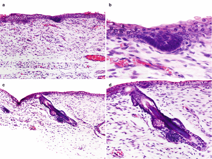
Apocrine glands are derived from the folliculo-sebaceous-apocrine germ, which consists of a small cluster of columnar epithelial cells whose nuclei appear crowded and with the peripheral layer arranged in palisade that appears on the undersurface of the primitive ectoderm during the third month of gestation (Fig. ). That aggregation of germinative cells assumes the shape of a crescent and resides immediately above a nubbin of mesenchymal cells. The cluster of germinative cells will give rise to the entire folliculo-sebaceous-apocrine unit, and the cluster of mesenchymal cells is devoted to become the future follicular papilla. During the next weeks of embryonic life, the folliculo-sebaceous-apocrine germ grows downward as a solid epithelial column that penetrates in an oblique way into the primitive dermis. Around the fifth month of embryonic life, along the side of a developing follicle that forms an obtuse angle with the primitive ectoderm, there are three discrete bulges, that, from the uppermost to the lowest one, are the bulge that becomes an apocrine gland and duct, the bulge that eventuates in a sebaceous gland and duct, and the bulge that acts as point of attachment for the fascicles of the arrector pili muscle and contains the follicular epithelial stem cells. Initially, the apocrine bulge develops in all follicles, but later it involutes in most of them, except in those of the so-called apocrine areas of the skin (see below).
Fig 11 Embryonic development of folliculo-sebaceous-apocrine unit a - photo 1
Next page
Light

Font size:

Reset

Interval:

Bookmark:

Make

Similar books «Cutaneous Adnexal Neoplasms»

Look at similar books to Cutaneous Adnexal Neoplasms. We have selected literature similar in name and meaning in the hope of providing readers with more options to find new, interesting, not yet read works.


Reviews about «Cutaneous Adnexal Neoplasms»

Discussion, reviews of the book Cutaneous Adnexal Neoplasms and just readers' own opinions. Leave your comments, write what you think about the work, its meaning or the main characters. Specify what exactly you liked and what you didn't like, and why you think so.