Jong-Soo Lee - Primary Eye Examination: A Comprehensive Guide to Diagnosis
Here you can read online Jong-Soo Lee - Primary Eye Examination: A Comprehensive Guide to Diagnosis full text of the book (entire story) in english for free. Download pdf and epub, get meaning, cover and reviews about this ebook. year: 2019, publisher: Springer, genre: Children. Description of the work, (preface) as well as reviews are available. Best literature library LitArk.com created for fans of good reading and offers a wide selection of genres:
Romance novel
Science fiction
Adventure
Detective
Science
History
Home and family
Prose
Art
Politics
Computer
Non-fiction
Religion
Business
Children
Humor
Choose a favorite category and find really read worthwhile books. Enjoy immersion in the world of imagination, feel the emotions of the characters or learn something new for yourself, make an fascinating discovery.

- Book:Primary Eye Examination: A Comprehensive Guide to Diagnosis
- Author:
- Publisher:Springer
- Genre:
- Year:2019
- Rating:3 / 5
- Favourites:Add to favourites
- Your mark:
Primary Eye Examination: A Comprehensive Guide to Diagnosis: summary, description and annotation
We offer to read an annotation, description, summary or preface (depends on what the author of the book "Primary Eye Examination: A Comprehensive Guide to Diagnosis" wrote himself). If you haven't found the necessary information about the book — write in the comments, we will try to find it.
Jong-Soo Lee: author's other books
Who wrote Primary Eye Examination: A Comprehensive Guide to Diagnosis? Find out the surname, the name of the author of the book and a list of all author's works by series.


Primary Eye Examination: A Comprehensive Guide to Diagnosis — read online for free the complete book (whole text) full work
Below is the text of the book, divided by pages. System saving the place of the last page read, allows you to conveniently read the book "Primary Eye Examination: A Comprehensive Guide to Diagnosis" online for free, without having to search again every time where you left off. Put a bookmark, and you can go to the page where you finished reading at any time.
Font size:
Interval:
Bookmark:

This Springer imprint is published by the registered company Springer Nature Singapore Pte Ltd.
The registered company address is: 152 Beach Road, #21-01/04 Gateway East, Singapore 189721, Singapore
The visual acuity test is the best way to show the status of eye. It includes uncorrected and corrected visual acuity, distant and near visual acuity, binocular and monocular visual acuity, decimal and fractional visual acuity, visual acuity measured with one and parallel object, logMAR (minimal angle of resolution) visual acuity, and central and out-of-central visual acuity. In general, visual acuity is the central visual acuity when viewed from the foveola. The visual acuity in ophthalmology refers to the corrected visual acuity which is the corrected visual acuity measured by refraction. If the uncorrected visual acuity is poor and the corrected visual acuity is good, the refractive error should be considered. If the corrected visual acuity is bad, a pathology from the eye to the central nervous system should be considered. The appropriate visual acuity test should be selected and recorded according to the subject and purpose. If the patient has the history of using contact lenses, cataract surgery, or LASIK surgery, record them on your visual acuity chart.
It is possible to examine at 3 years of age or older. At 38 years of age, the visual acuity is measured higher when using single object chart than the parallel chart. That is, all persons who can read the target in the visual acuity chart are all candidates.
The causes of visual impairment include refractive errors and eye diseases involving visual pathway. The refractive error can be improved by correction, but for the cases of eye diseases, it is difficult to improve the visual acuity only by correcting the refractive error. Therefore, visual acuity test can be a great help in determining the cause of vision impairment.
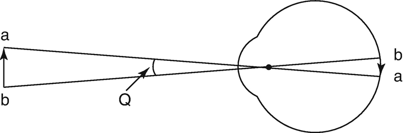
The reciprocal of the minimum visual angle is visual acuity. If the visual angle Q is 2 minutes, the visual acuity is 0.5
The visual acuity chart includes a decimal visual acuity chart, a fractional visual acuity chart (Snellen visual acuity chart), and a logMAR chart. The devices of the visual acuity chart include reflection type, transmission type, and projection type.
The decimal visual acuity is 0.1 intervals from 0.1 to 1.0 and 1.2, 1.5, and 2.0 afterward. Because it is expressed as a reciprocal of the minimum visual angle, the arrangement does not show the same interval. 0.1 and 0.2 and 0.9 and 1.0 are both one step, but the visual angle is 10 minutes and 5 minutes in the former and 1.1 minutes and 1 minute in the latter. The former is twice the difference, and the latter is 1.1 times, which does not represent the same difference. Therefore, if 0.125 and 0.16 are inserted between 0.1 and 0.2, 0.25 is inserted between 0.2 and 0.3, and 0.7 and 0.9 are subtracted, the arrangement of decimal visual acuity chart becomes almost equal.
The fractional visual acuity (Snellen method) has the same arrangement of visual acuity, and unlike the decimal visual acuity charts, it is possible to indicate by two-step visual acuity increase or decrease.
The logarithmic visual acuity chart displays the visual acuity in logMAR, which is the logarithm of the minimal angle of resolution (MAR). Each step of the visual acuity chart is at the same interval, so an arithmetic average is possible to be calculated.
The logarithmic visual acuity chart has five characters per line, and the left and right character spacing is a spacing of the size of each character, and the size ratio of the large character and the lower character is kept at a constant ratio of about 5:4, respectively. The spacing between the upper and lower lines is the size of the character in the lower row. They are arranged at the same interval at which the index is reduced by 0.1 by one logMAR at the top. This visual acuity chart is used at a distance of 4 m. It can measure up to 20/957, that is, 0.02 at 1 m, which is useful for patients with low vision.
Font size:
Interval:
Bookmark:
Similar books «Primary Eye Examination: A Comprehensive Guide to Diagnosis»
Look at similar books to Primary Eye Examination: A Comprehensive Guide to Diagnosis. We have selected literature similar in name and meaning in the hope of providing readers with more options to find new, interesting, not yet read works.
Discussion, reviews of the book Primary Eye Examination: A Comprehensive Guide to Diagnosis and just readers' own opinions. Leave your comments, write what you think about the work, its meaning or the main characters. Specify what exactly you liked and what you didn't like, and why you think so.