Craig A. Canby - Problem-Based Anatomy, 1e
Here you can read online Craig A. Canby - Problem-Based Anatomy, 1e full text of the book (entire story) in english for free. Download pdf and epub, get meaning, cover and reviews about this ebook. City: Philadelphia, year: 2006, publisher: Elsevier Health Sciences;Saunders, genre: Children. Description of the work, (preface) as well as reviews are available. Best literature library LitArk.com created for fans of good reading and offers a wide selection of genres:
Romance novel
Science fiction
Adventure
Detective
Science
History
Home and family
Prose
Art
Politics
Computer
Non-fiction
Religion
Business
Children
Humor
Choose a favorite category and find really read worthwhile books. Enjoy immersion in the world of imagination, feel the emotions of the characters or learn something new for yourself, make an fascinating discovery.

- Book:Problem-Based Anatomy, 1e
- Author:
- Publisher:Elsevier Health Sciences;Saunders
- Genre:
- Year:2006
- City:Philadelphia
- Rating:5 / 5
- Favourites:Add to favourites
- Your mark:
Problem-Based Anatomy, 1e: summary, description and annotation
We offer to read an annotation, description, summary or preface (depends on what the author of the book "Problem-Based Anatomy, 1e" wrote himself). If you haven't found the necessary information about the book — write in the comments, we will try to find it.
- Features more than 80 clinical scenarios that promote interactive learning and build a foundation of knowledge for clinical practice.
- Presents information in seven sections to correspond with a regional approach to anatomy: head and neck, back, thorax, pelvis and perineum, upper extremity, and lower extremity.
- Covers the subdisciplines of anatomy including anatomic pathology cell biology embryology gross anatomy histology neuroanatomy and radiologic anatomy.
- Includes references to Grays Anatomy for Students, and follows a parallel organization, making it easy to use both books together.
Craig A. Canby: author's other books
Who wrote Problem-Based Anatomy, 1e? Find out the surname, the name of the author of the book and a list of all author's works by series.

Problem-Based Anatomy, 1e — read online for free the complete book (whole text) full work
Below is the text of the book, divided by pages. System saving the place of the last page read, allows you to conveniently read the book "Problem-Based Anatomy, 1e" online for free, without having to search again every time where you left off. Put a bookmark, and you can go to the page where you finished reading at any time.
Font size:
Interval:
Bookmark:
PROBLEM-BASED Anatomy
Craig A. Canby, Ph.D.
Associate Professor of Anatomy, Coordinator of University Accreditation, Des Moines UniversityOsteopathic Medical Center, Des Moines, Iowa
SAUNDERS
Front Matter
Craig A. Canby, Ph.D.
Associate Professor of Anatomy
Coordinator of University Accreditation
Des Moines UniversityOsteopathic Medical Center
Des Moines, Iowa
Copyright
SAUNDERS ELSEVIER
1600 John F. Kennedy Boulevard
Suite 1800
Philadelphia, PA 19103-2899
Problem-Based Anatomy
Copyright 2006 by Elsevier Inc. All rights reserved.
ISBN 13: 978-1-4160-2417-0
ISBN 10: 1-4160-2417-4
), by selecting Customer Support and then Obtaining Permissions.
NOTICE
Knowledge and best practice in this field are constantly changing. As new research and experience broaden our knowledge, changes in practice, treatment and drug therapy may become necessary or appropriate. Readers are advised to check the most current information provided (i) on procedures featured or (ii) by the manufacturer of each product to be administered, to verify the recommended dose or formula, the method and duration of administration, and contraindications. It is the responsibility of the practitioner, relying on his or her own experience and knowledge of the patient, to make diagnoses, to determine dosages and the best treatment for each individual patient, and to take all appropriate safety precautions. To the fullest extent of the law, neither the Publisher nor the Editor assumes any liability for any injury and/or damage to persons or property arising out or related to any use of the material contained in this book.
Library of Congress Cataloging-in-Publication Data
Canby, Craig A.
Problem-based anatomy / Craig A. Canby
p. ; cm.
ISBN 1-4160-2417-4
1. Human anatomyProblem, exercises, etc. 2. Human anatomyCase studies. 3. Physiology, PathologicalProblems, exercises, etc. 4. Physiology, PathologicalCase studies. I. Title.
[DNLM: 1. AnatomyCase Reports. 2. AnatomyProblems and Exercises. QS 18.2 C214p 2006]
QM32.C36 2006
611.0076dc22
2005042680
Acquisitions Editor: William Schmitt
Development Editor: Kevin Kochanski
Senior Project Manager: Mary Stermel
Marketing Manager: John Gore
Printed in China.
Last digit is the print number: 9 8 7 6 5 4 3 2 1
Dedication
To my family for their blessings of love, support, and patience, and to my students who have blessed my professional life.
PREFACE
Over the years, the curricula of health professions programs have evolved into a state of integration. From the first day of school, health professional students are immersed in a curriculum that exposes them to the basic sciences and the clinical sciences. This curricular shift has also impacted instruction in the discipline of anatomy. While anatomists remain passionate about the noble act of transferring the purity of anatomical knowledge onto future generations of students, they are also mindful that students in the health professions require a different fund of anatomical knowledge. These students must be able to apply their anatomical knowledge base to clinical situations. In response, anatomy educators are clearly emphasizing the clinical relevance of anatomy in their education of future generations of health professional students.
This text was written to further the clinical significance of anatomy. The text features 61 clinical cases divided into the traditional seven regions of the body: 1) back, 2) thorax, 3) abdomen, 4) pelvis and perineum, 5) lower limb, and 6) upper limb, and 7) head and neck. Each clinical case frames a set of study questions and accompanying answers. Another value-added attribute of this format is its integrated approach to the broad field of anatomy. Under the umbrella of anatomy are the subdisciplines of anatomic pathology, cell biology, embryology, gross anatomy, histology, neuroanatomy, and radiologic anatomy. The questions accompanying each clinical case are crafted, wherever possible, to include these anatomical subdisciplines. Additionally, 185 multiple choice questions are provided in Section VIII so that the student can assess his/her mastery of the material. Answers to the questions and references back to the text are also provided.
This text can be used by students as a clinically-oriented complement to standard anatomy-related texts in their courses. It should also be of educational utility for students preparing for national licensing examinations and to residents who are in the early years of their graduate medical training.
As with any intellectual pursuit, it is always proper to gratefully acknowledge the contributions that others have made to the outcome. To that end, I offer my profound gratitude to the authors of other textbooks who graciously granted permission to include their figures in this book. The textbooks are Grays Anatomy for Students by Drake et al, The Developing Human, 7th edition by Moore and Persaud, Sabiston Textbook of Surgery, 17th edition by Townsend et al, Robbins and Cotran Pathologic Basis of Disease, 7th edition by Kumar et al, and Color Textbook of Histology, 2nd edition by Gartner and Hiatt. The student who desires a greater depth of knowledge in the topics covered in this book is encouraged to use these outstanding resources for additional information.
Anatomy is a wonderful, noble discipline filled with the awe and wonders of human form. It is a privilege and a blessing to be touched by the sanctity of its incredible beauty. I hope you are rewarded with the teachings of anatomy in your professional education and practice as I have been in my professional career. As learning colleagues of anatomy, please contact me directly with constructive criticisms about this text for incorporation into future editions.
CRAIG A. CANBY
ACKNOWLEDGMENTS
I am profoundly grateful to the many publication professionals who have played pivotal roles in shepherding this book through its preparation and production. Through their genuine diligence, creativity, and a keen eye to detail they have transformed a concept for a book and later a black and white manuscript into a vibrant and superbly edited publication. My special thanks go to William Schmitt for the opportunity to author this book and to Kevin Kochanski for his developmental acumen.
SECTION I
BACK
CASE 1
).
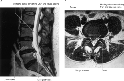
FIGURE 1-1 MRI of a herniated disc between L4 and L5 vertebrae. A, Sagittal plane. B, Axial plane.
(Drake R, Vogl W and Mitchell A: Grays Anatomy for Students. Churchill Livingstone, 2004. Fig. 2-33.)
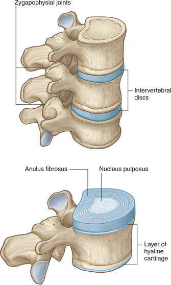
). A disc is not found between the atlas and the axis. The discs are thinnest between the cervical vertebrae and progressively thicken as they descend the vertebral column. In the secondary curvatures of the vertebral column (cervical and lumbar regions), the intervertebral discs are thicker anteriorly. This produces the anterior convexity of the cervical and lumbar segments of the vertebral column.
Font size:
Interval:
Bookmark:
Similar books «Problem-Based Anatomy, 1e»
Look at similar books to Problem-Based Anatomy, 1e. We have selected literature similar in name and meaning in the hope of providing readers with more options to find new, interesting, not yet read works.
Discussion, reviews of the book Problem-Based Anatomy, 1e and just readers' own opinions. Leave your comments, write what you think about the work, its meaning or the main characters. Specify what exactly you liked and what you didn't like, and why you think so.