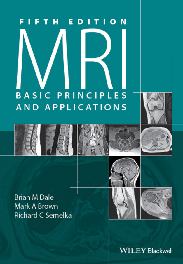
This edition first published 2015 2015 by John Wiley & Sons, Ltd
Registered office: John Wiley & Sons, Ltd, The Atrium, Southern Gate, Chichester, West Sussex, PO19 8SQ, UK
Editorial offices: 9600 Garsington Road, Oxford, OX4 2DQ, UK
111 River Street, Hoboken, NJ 07030-5774, USA
For details of our global editorial offices, for customer services and for information about how to apply for permission to reuse the copyright material in this book please see our website at www.wiley.com/wiley-blackwell
The right of the author to be identified as the author of this work has been asserted in accordance with the UK Copyright, Designs and Patents Act 1988.
All rights reserved. No part of this publication may be reproduced, stored in a retrieval system, or transmitted, in any form or by any means, electronic, mechanical, photocopying, recording or otherwise, except as permitted by the UK Copyright, Designs and Patents Act 1988, without the prior permission of the publisher.
Designations used by companies to distinguish their products are often claimed as trademarks. All brand names and product names used in this book are trade names, service marks, trademarks or registered trademarks of their respective owners. The publisher is not associated with any product or vendor mentioned in this book. It is sold on the understanding that the publisher is not engaged in rendering professional services. If professional advice or other expert assistance is required, the services of a competent professional should be sought.
The contents of this work are intended to further general scientific research, understanding, and discussion only and are not intended and should not be relied upon as recommending or promoting a specific method, diagnosis, or treatment by health science practitioners for any particular patient. The publisher and the author make no representations or warranties with respect to the accuracy or completeness of the contents of this work and specifically disclaim all warranties, including without limitation any implied warranties of fitness for a particular purpose. In view of ongoing research, equipment modifications, changes in governmental regulations, and the constant flow of information relating to the use of medicines, equipment, and devices, the reader is urged to review and evaluate the information provided in the package insert or instructions for each medicine, equipment, or device for, among other things, any changes in the instructions or indication of usage and for added warnings and precautions. Readers should consult with a specialist where appropriate. Further, readers should be aware that Internet Websites listed in this work may have changed or disappeared between when this work was written and when it is read. No warranty may be created or extended by any promotional statements for this work. Neither the publisher nor the author shall be liable for any damages arising herefrom.
Library of Congress Cataloging-in-Publication Data
Dale, Brian M., author.
MRI : basic principles and applications / Brian M. Dale, Mark A. Brown, and Richard C. Semelka. Fifth edition.
p. ; cm.
Preceded by MRI / Mark A. Brown, Richard C. Semelka. 4th ed. c2010.
Includes bibliographical references and index.
ISBN 978-1-119-01305-1 (pbk.)
I. Brown, Mark A., 1955- , author. II. Semelka, Richard C., author. III. Brown, Mark A., 1955- MRI. Preceded by (work): IV. Title.
[DNLM: 1. Magnetic Resonance Imaging--methods. WN 185]
RC78.7.N83
616.07548dc23
2015015322
A catalogue record for this book is available from the British Library.
Wiley also publishes its books in a variety of electronic formats. Some content that appears in print may not be available in electronic books.
Preface
Magnetic Resonance Imaging (MRI) continues to be an integral component of medical imaging. New measurement techniques and applications continue to be developed nearly thirty years after the initial clinical scanners were installed. Even so, the basic principles behind the measurement techniques remain as true today as then. This book was written to present the fundamental concepts of MRI in a clear and concise manner, minimizing the mathematical formalism yet providing a foundation to understand the results that are obtained with today's clinical scanners.
Since the fourth edition, parallel imaging and high channel-count coil arrays have become mainstream clinical tools. We have added material describing the physics behind these modern techniques. We have also added a focus on material that is relevant to radiologist board exams. Such material is highlighted in the text and will now be easier to find and identify. It is interspersed with other material that remains important for a deeper understanding of the physics of MRI and provides additional clarity. New and updated figures are included throughout the book.
As always, many people must be thanked for their help in this. First, we would like to thank the technical staff at the Siemens Training and Development Center and the faculty, fellows, and staff at the University of North Carolina for their interest in this project and their assistance in its completion. In addition, thanks to Wolfgang Rehwald, James R. MacFall, and H. Cecil Charles for providing images. Finally, thanks to our families for their support and patience in this project.
B.M. Dale
M.A. Brown
R.C. Semelka
ABR study guide topics
Where you see text within lines and accompanied by the  logo, this indicates content that would be especially useful for those studying for the American Boards of Radiology.
The list below indicates the location of that content throughout the book.
- Magnetic fields
- Magnetic susceptibility 1.6
- Types of magnetic materials 1.6
- Magnetic Fields (B) 1.1
- Magnetic moment interaction with an external field (B0): the Larmor equation and precessional frequency 1.4
- Net magnetization due to B0 and field strength 1.5
- Nuclear MR and excitation 2.1
- MR signal properties
- Spin density (proton) 3.1
- T2 (transverse) relaxation 3.2
- T2* relaxation 3.2
- T1 (longitudinal) relaxation 3.1
- T1 weighting, T2 weighting, proton density-weighting 6.1
- Pulse sequences and contrast mechanisms
- Echo time (TE), repetition time (TR), and inversion time (TI) 6.1, 6.4
- Spin-echo (SE) pulse sequences 6.1
- Inversion-recovery spin-echo pulse sequences 6.4
- Gradient-echo (GE or GRE) pulse sequences 6.2
- Echo-planar (EPI) pulse sequences 6.3
- Fast- or turbo-spin-echo (FSE) pulse sequences 6.1
- Manipulation of pulse sequence characteristics 7.1
- MR instrumentation
- Static magnetic field (B0) systems 14.2
- Gradient fields and the gradient subsystem 14.3
- Shimming and shim coils 14.2
- Radiofrequency transmitter (B1) subsystem 14.4
- Radiofrequency receiver subsystem 14.5
- Radiofrequency coils 14.5
- Spatial localization
- Slice-selection 4.2
- Phase-encoding 4.4
- Frequency-encoding 4.3
- Two-dimensional Fourier transform (2DFT) image reconstruction 5.3
- k-space description 5.5
- Methods of filling k-space 5.6, 5.7
- Image characteristics
- Factors affecting spatial resolution 4.2, 4.3, 4.4
- Factors affecting signal-to-noise ratio (SNR) 5.4, 7.3.2
Next page




 logo, this indicates content that would be especially useful for those studying for the American Boards of Radiology.