1.1 Anatomy of the Anorectum
Anal fissure (Fissure-in-ano) becomes manifest at the caudal end of the anatomical anal canal. Therefore, it is necessary to explain the compartments forming the anal canal. Also, neural structures must be dealt with, because an increased sphincter muscle tone often assumed to be the origin of an anal fissure could then be explained as the consequence of a disturbed sequence of reflex actions.
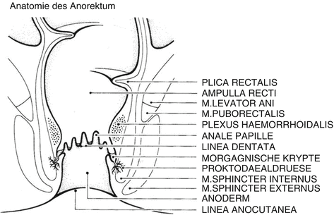
In evolutionary terms, ectoderm and endoderm adjoin each other in the anal canal. The so-called anatomical anal canal is lined by ectoderm and ends at the level of the pectinate line, which can be macroscopically identified by the indentations of the anal crypt line. Thus, the anatomical anal canal is defined topographically. The so-called surgical anal canal additionally comprises the endodermal portion of the anal canal proximal to the anatomical anal canal up to the level of the anorectal ring. There, the anal canal widens to become the rectal ampulla. The anorectum guarantees the continence function of the colon.
Anorectal Muscles
The muscular portions of the continence organ wrap around the anal canal most likely comparable to two tubes, one inside the other. The inner tube consists of the internal sphincter muscle as the continuation of the circular musculature of the rectum. This is smooth musculature which is due to colonic aganglionosis capable of permanent contraction, thus keeping the anal canal tightly closed. Pressure measurements show that it accounts for about 70% of the muscle tone at rest. Under spinal anesthesia with a corresponding elimination of the striated muscle portions, the resting pressure profile of the anal canal remains largely unchanged. This underlines the importance of the internal anal sphincter muscle as the essential continence factor. On the other hand, it makes defecation possible due to its relaxation reflex when the rectal ampulla is distended.
The striated external anal sphincter muscle encloses the internal anal sphincter muscle at the caudal end of the anal canal. Similar to the striated skeletal musculature, the external sphincter muscle is capable of a maximal short-period contraction for the purpose of deliberate action. Cranially, the striated puborectalis muscle follows, which genetically corresponds to the lowest part of the levator ani muscle. It encases dorsally the rectum like a sling, while omitting its ventral parts and inserting into the pubic bone (os pubis) with broad tendons. The contraction of this muscle leads to a diagonal sealing of the upper anal canal by a ventral shift of the back wall of the lower rectum like a crimp connection (gross anal closure).
Epithelial Portions of the Continence Organ
The epithelial coating of the anorectum consists of three parts. The epithelium of the distal part of the anal canal, the anatomical anal canal, is especially important for the continence function. Here lies the dry, nonkeratinizing squamous epithelium of the anoderm, which due to its intensive supply with sensitive nerve endings is capable to differ between the various aggregate conditions of the colonic contents. Above the pectinate line, in the section of the surgical anal canal stemming from the endoderm, a columnar epithelium is found whose structure is similar to that of the rectal mucosa. Between these two areas lies the zone of the so-called transitional epithelium (transitional cell area), which is of individually differing width and is also supplied with sensitive nerves. The histological examination discloses a cylindrical structure. Macroscopically, the transitional cell area shows a whitish color in contrast to the reddish shimmering mucosa above, which is not innervated sensitive.
Subepithelial Portions of the Continence Organ
Under the mucosa of the upper anal canal, the corpus cavernosum recti is located immediately below the epithelium. It is in fact a set of venous vascular cushions which exist in every individual and are colloquially called hemorrhoids (Aigner et al. ). The arterial supply of this vascular plexus takes place cranially in the tunica submucosa, in particular through the branches of the superior rectal artery (A. rectalis superior). The venous discharge runs submucosally partly into the superior rectal vein (V. rectalis superior) and partly into the venous stems of the middle rectal veins (Vv. rectalis mediae), while taking its course through the internal anal sphincter. The virtually permanent contraction of this muscle to maintain continence leads to a partial obstruction of the venous discharge, while the arterial supply remains undisturbed. As a result, the vascular cushions are dammed up, and thus seal effectively the lumen of the upper anal canal (hermetic seal). The caudal portions of the branches of the superior rectal artery pass through the muscular rectal wall in a straight direction and reach the corpus cavernosum recti without taking its course through the submucosa. Additionally, there is arterial supply of the corpus cavernosum recti from the middle rectal artery (A. rectalis media), so that this redundant vascularization ensures the function of the corpus cavernosum under all circumstances.
Neural Portions of the Continence Organ
The neural structures make sure that the different functional areas of the continence organ are connected with each other and also with the superordinate nerve centers (Raulf ).
Fig. 1.1
Anorectal anatomy, schematic (Wienert and Mlitz )
1.2 Definition of Anal Fissure
Anal fissures are divided into primary acute or chronic and secondary fissures. They are lesions in the lower anal canal. Primary anal fissures develop as local entities; secondary fissures develop in the context of a basic disease.
Anal fissure is a painful, linear or spindle-shaped, longitudinal defect in the highly sensitive anoderm. It often causes a reactive sphincter hypertonia, which possibly triggers a reduced blood circulation with venous stasis. It is found in the anoderm between the pectinate line and the anal verge.
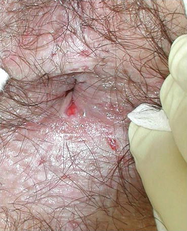
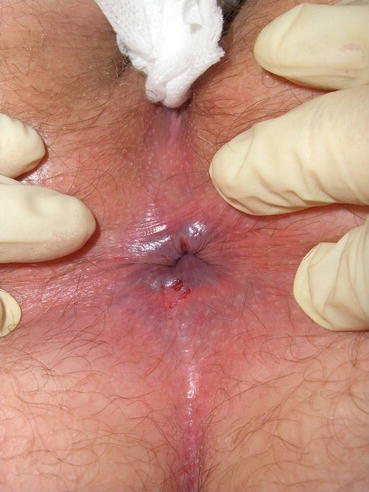
Anal fissures can heal either spontaneously or after therapy. They can recur or turn from acute into chronic anal fissures (Figs. ).
Fig. 1.2
Acute anal fissure dorsal, erosions perianal
Fig. 1.3
Acute anal fissure ventral, erosions dorsal

