Springer Science+Business Media LLC 2017
Puay Hoon Tan and Aysegul A. Sahin Atlas of Differential Diagnosis in Breast Pathology Atlas of Anatomic Pathology 10.1007/978-1-4939-6697-4_1
1. Normal Breast and Physiological Changes
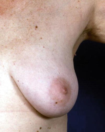
The breast is a modified sweat gland located in the superficial fascia of the anterior chest wall. The mature female breast has a distinctive protuberant, mound-shaped, or conical form and covers the area from the second or third rib to the sixth or seventh rib (Fig. ).
The breast is made up of glandular and ductal elements embedded within fibrofatty tissue with a ratio of glandular to fibrofatty tissue that varies among individuals (Fig. ]. The largest amount of breast parenchyma is located in the upper outer quadrant, where the majority of cancers develop. An axillary tail of breast tissue often extends into the axilla. Before puberty, female and male breasts have the same appearance. The structure of the breast is under the influence of hormones, growth, and differentiation factors. When puberty begins in females, mammary ducts branch out, terminal duct buds are formed, and the stromal component (mainly adipose tissue) of the breast proliferates. Both stroma and epithelium undergo changes during the menstrual cycle, pregnancy, lactation, and menopause. During male puberty, breast development is limited to rudimentary large duct development without breast enlargement.
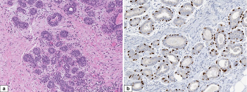
The mammary epithelium is ectodermally derived. Small segments of lactiferous duct orifices at the nipple are lined by squamous epithelium, while the rest of the breast ductal system is lined by two cell layers, inner luminal cells and outer myoepithelial cells, surrounded by the basement membrane (Fig. ). The intralobular stroma is usually loose and more cellular than the interlobular stroma, and unlike the interlobular stroma, it usually does not have adipose tissue. Intralobular stroma is hormone sensitive and shows cyclic histologic changes.
Fig. 1.1
Normal adult female breast. Photograph of a normal female prior to undergoing prophylactic mastectomy
Fig. 1.2
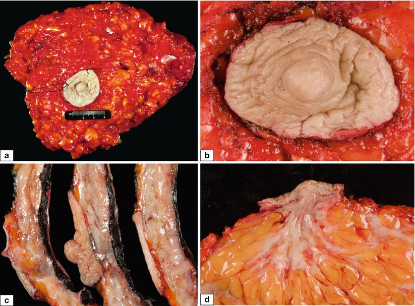
Normal adult female breast. Gross features. ( a ) Skin-sparing total mastectomy specimen showing full extension of breast tissue with the nipple located in the normal central position. ( b ) Higher magnification of the nipple-areolar complex. ( c ) Cut section through the nipple shows dense subareolar fibrous connective tissue. ( d ) Higher magnification of ( c ) showing subareolar fibrous tissue radiating into the fatty breast parenchyma
Fig. 1.3
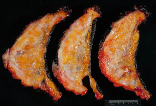
Normal adult female breast. Gross features. Cut sections of breast mastectomy specimen. The ratio of fat to fibrous tissue is variable and correlates with mammographic density
Fig. 1.4
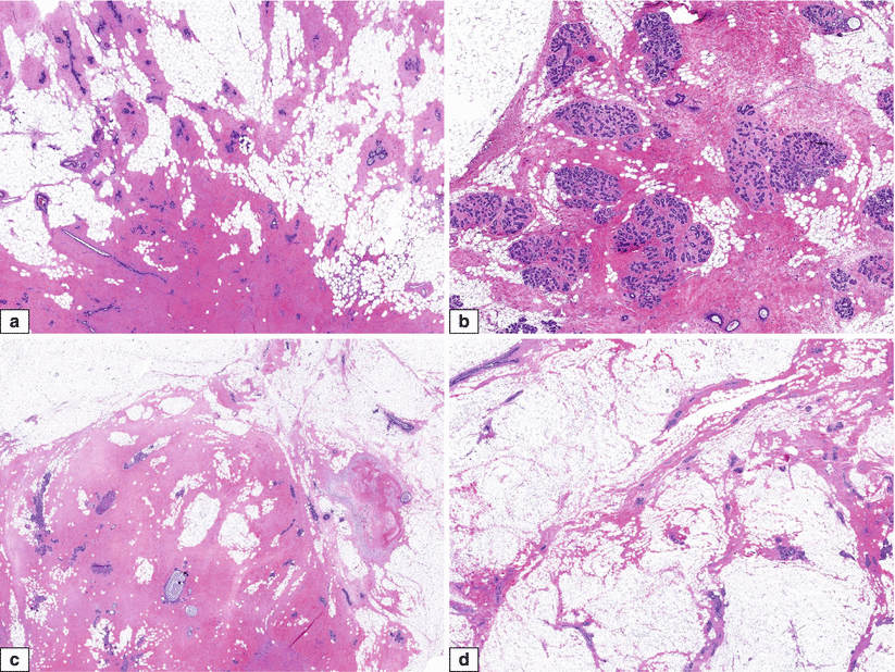
Normal adult female breast. Histologic features. ( ad ) H&E sections showing varying ratios of fat to fibrous tissue in stroma and various amounts of glandular elements from different cases
Fig. 1.5
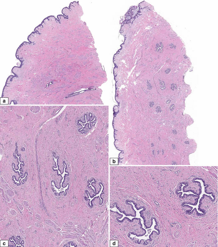
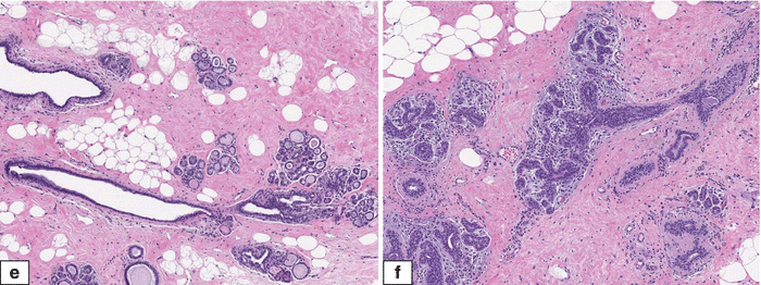
Normal adult female breast. Histologic features. ( ad ) H&E sections of the nipple-areolar complex showing nipple or lactiferous ducts extending from the skin surface into the breast parenchyma. The lactiferous ducts show a branching shape and are lined by bilayered epithelium (luminal epithelial and outer myoepithelial cells). There may be stromal folds protruding into the ductal lumens which should not be mistaken for a papillary lesion. Smooth muscle bundles are seen among the lactiferous ducts. ( e , f ) Terminal ductal lobular unit, which is the functional unit of breast parenchyma, consists of a feeding duct with branching acini embedded in connective tissue
Fig. 1.6
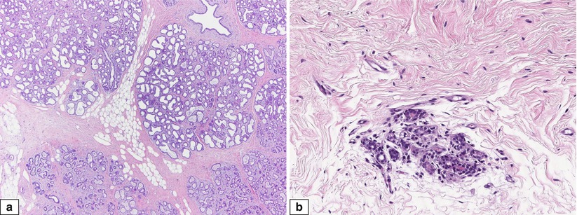
Normal adult female breast with physiological changes. Histologic features. ( a ) Expanded terminal ductal lobular unit of a lactating breast comprises an increased number of acini with luminal secretions. ( b ) Atrophic terminal ductal lobular unit of a postmenopausal female
Fig. 1.7
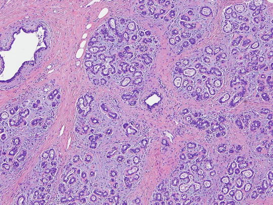
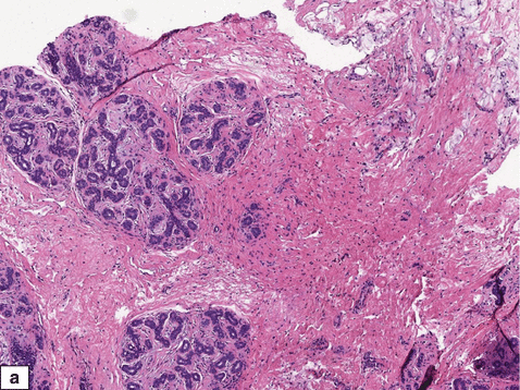
Normal adult female breast. Histologic features. Intralobular stroma shows a loose myxoid appearance, with less collagenisation compared to interlobular fibrous tissue
Fig. 1.8
Normal adult female breast. Histologic and immunohistochemical features. ( a ) A lobule composed of multiple acini. The luminal epithelial cells are surrounded by myoepithelial cells, some of which have clear cytoplasm. ( b ) Immunohistochemical staining for p63 highlights myoepithelial cell nuclei
Fig. 1.9
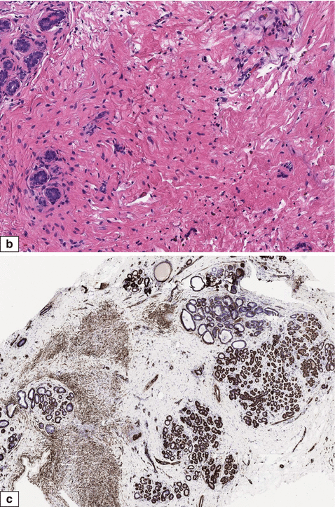
Normal adult female. ( a , b ) Stroma shows myoid cells in addition to fibroblasts. ( c ) Spindle cells with myoid differentiation show immunoreactivity for smooth muscle myosin
Physiological Changes
Menstrual Cyclic Changes
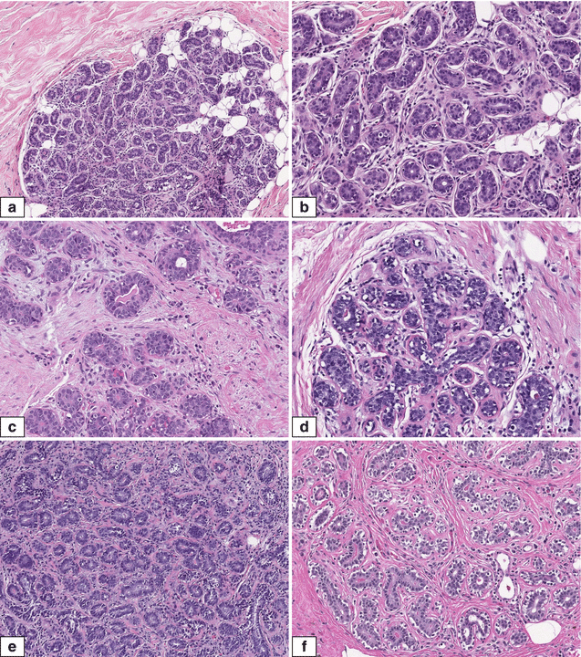
Both stromal and glandular components of the breast undergo histologic changes during the menstrual cycle. However, these changes are not distinct and specific, unlike changes observed in endometrial epithelium during the menstrual cycle [].
Fig. 1.10
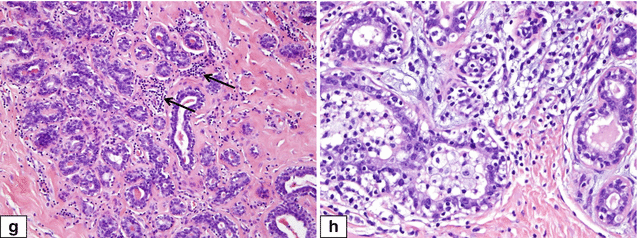
Normal adult female breast. Histologic features of menstrual cycle phases. ( a ) Proliferative phase: H&E section shows lobular unit composed of tightly packed acini. ( b ) Acini are lined by crowded, poorly oriented cells with little or no lumen formation. No secretion is evident. The intralobular stroma is dense and cellular. ( c ) Follicular phase: H&E shows cells lining the acini becoming columnar with central lumens that are apparent. ( d ) Lumens have minimal secretions. ( e ) Secretory phase: Acini have open lumens which contain secretions. ( f ) The lobular stroma is loose. Both epithelial and myoepithelial cells are distinct. ( g ) Closing of the cycle: Small acini associated with dense stroma which contains inflammatory cells ( arrows ). ( h ) Higher magnification shows distinct clear-cell appearances of peripheral myoepithelial cells

