Bianca Maria Piraccini Nail Disorders 2014 A Practical Guide to Diagnosis and Management 10.1007/978-88-470-5304-5_1
Springer-Verlag Italia 2014
1. Nail Anatomy and Physiology for the Clinician
Abstract
The nail plate is the final product of proliferation and differentiation of nail matrix keratinocytes. It emerges from underneath the proximal nail fold and grows distally at a median speed of 13 mm/month. The nail plate is firmly attached to the nail bed and detaches at the hyponychium, a small transverse band of epithelium that is in continuity with the pulp. Laterally, it is lined by the lateral nail folds. Each nail constituent has a role in production and growth of a healthy nail plate and if damaged produces typical nail symptoms: nail matrix damage will impair nail plate appearance and growth and nail bed and hyponychium damage with impair nail plate adhesion to the underlying tissues.
Examination of the nail should therefore include all parts of the nail apparatus: proximal and lateral folds, nail bed and hyponychium, and nail plate. The nail matrix is not visible directly, but only its distal part can be seen under the proximal nail plate and a distally convex crescent: the lunula.
The nails have several important uses, which are easily appreciable when the nails are absent or they lose their function. The most evident use of fingernails is to be an ornament of the hand, but we must not underestimate other important functions, such as the protective value of the nail plate against trauma to the underlying distal phalanx, its counterpressure effect to the pulp important for walking and for tactile sensation, the scratching function, and the importance of fingernails for manipulation of small objects.
The nails can also provide information about the persons work, habits, and health status, as several well-known nail features are a clue to systemic diseases. Abnormal nails due to biting or onychotillomania give clues to the persons emotional/psychiatric status. Nail samples are utilized for forensic and toxicology analysis, as several substances are deposited in the nail plate as they are produced and remain stored during growth.
It is therefore important to know how the healthy nail appears and how it is formed, in order to detect signs of pathology and understand their pathogenesis.
1.1 Nail Anatomy and Physiology
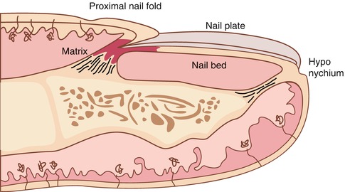
What we call nail is the nail plate, the final part of the activity of 4 epithelia that proliferate and differentiate in a specific manner, in order to form and protect a healthy nail plate [) is composed by:
Fig. 1.1
Drawing of the nail apparatus in transverse section, showing the four structures that contribute to nail plate formation and growth: proximal nail fold, nail matrix, nail bed, and hyponychium. Note the proximity of the nail apparatus with the bone of the distal phalanx and the two ligaments that link them together
Nail matrix: responsible for nail plate production
Nail folds: responsible for protection of the nail matrix
Nail bed: responsible for nail plate adhesion during growth
Hyponychium: responsible for nail plate detachment at the distal margin of the digit
1.2 Nail Plate
The nail plate is a fully keratinized, dead structure, which is continuously produced by the nail matrix . Nail matrix keratinocytes proliferate and undergo a sudden differentiation with loss of nuclei and strict intercellular adherence that give rise to the nail plate. The plate emerges from underneath the proximal nail fold and grows distally, surrounded by the lateral folds and adherent to the nail bed , until it detaches in correspondence to the hyponychium .
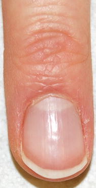
The nail plate has a rectangular shape, is semitransparent, and has a smooth shiny surface, lined by thin longitudinal lines that increase with ageing (Fig. ). It appears pink when attached to the nail bed, as it allows visualization of nail bed blood vessels, while the free edge is whitish in color. Proximally, the nail plate of fingernails and of some toenails shows a distally convex white area, the lunula , which corresponds to the distal nail matrix. The shape of the lunula determines the shape of the distal margin of the nail plate. Before the nail plate free margin, there is a transverse reddish band (the onychodermal and onychocorneal bands) that corresponds to the nail isthmus , the area of the strongest adherence between nail plate and nail bed.
Fig. 1.2
Normal fingernail: the plate emerges from the proximal nail fold, lined by the cuticle. The plate color is pink, with a proximal oval whitish area, the lunula. The free margin is white
The nail plate physical characteristics are unique and essential for its uses: it is hard and difficult to break, but elastic and bendable, resistant to chemicals, and strictly adherent to the underlying tissues.
These features are due to its high content in hard keratins, which constitute its 8090 %, and to its particular anatomical structure that includes three layers:
The dorsal part, 0.080.1 mm thick, consisting of tight, flattened cells, whose keratin filaments are oriented parallel and perpendicular to the growth axis. This portion gives the nail hardness and sharpness and is produced by the proximal nail matrix.
The intermediate nail plate, 0.30.5 mm thick, consisting of wide and irregular cells, with keratin perpendicular to the growth axis. This portion gives the nail flexibility and elasticity.
The ventral nail plate, 0.060.08 mm thick, produced by the nail bed, is necessary for the adhesion of the nail plate to the nail bed.
1.3 Nail Matrix
The matrix produces the nail plate continuously throughout life. It lies well protected under the proximal nail fold and just above the bone of the distal phalanx, to which is connected by a tendon that reaches the distal interphalangeal joint (Fig. ).
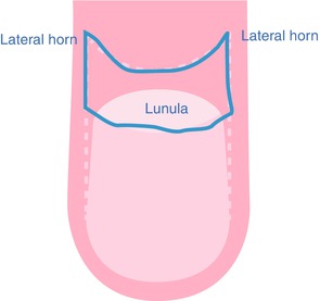
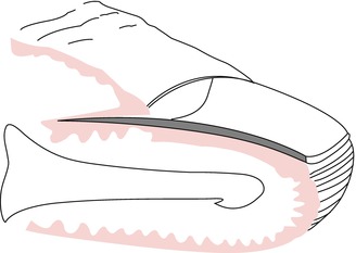
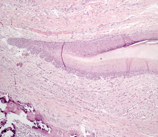
In order to understand the anatomy of the matrix, we should look at it both in a frontal and in transverse view. In frontal view (Fig. ). Matrix cells lose their nuclei and are strictly adherent to each other, with a cytoplasm completely filled by hard keratins. This gives rise to a completely transparent, flexible but hard, and resistant nail plate.
Fig. 1.3
Drawing of the matrix in frontal view: it has a horseshoe shape with distal convexity and two lateral horns
Fig. 1.4
Transverse section of the nail, showing the different epithelia. The matrix has a v-shaped appearance and distally continues with the nail bed

