Part I
Basic Concepts, Mechanisms of Disease and Pre-operative Planning
Mayo Foundation for Medical Education and Research 2017
Gustavo S. Oderich (ed.) Endovascular Aortic Repair 10.1007/978-3-319-15192-2_1
1. Historical Aspects and Evolution of Fenestrated and Branched Technology
Blayne Roeder 1
(1)
Aortic Intervention, Cook Medical, 750 Daniels Way, Box 489, Bloomington, IN 47402-0489, USA
(2)
Radiology Department, Royal Perth Hospital, 73 Sandpiper Island, Wannanup, WA, 6210, Australia
(3)
Faculty of Health Sciences, Curtin University of Technology, Bentley Campus, Perth, WA, 6845, Australia
Blayne Roeder (Corresponding author)
Email:
Michael Lawrence-Brown
Email:
Keywords
Abdominal Thoracoabdominal Arch Endovascular Aneurysm Repair Visceral Branch Fenestration Development
Introduction
Endovascular repair of aortic aneurysms (EVAR) has been disseminated worldwide since the first report by Juan Parodi in 1991 (Fig. ). Since this initial disclosure, several innovators have worked on broadening the indications of EVAR to treat patients with complex anatomy. The Zenith AAA Endovascular Graft resulted from the collaboration of a global team of innovators. This team shared a common philosophy that the endovascular repair must seal in healthy aorta to provide a durable repair over the life of the patient. As a result of this philosophy, the Zenith was the first endovascular graft to incorporate proximal fixation and a modular three-piece system in combination with a delivery system that allowed for precise deployment. This enables the physician to place the graft in a position that takes advantage of all the available healthy aortic seal zone possible, while active fixation acts to maintain the seal over a prolonged period, ultimately resulting in a durable endovascular repair. Subsequently, nearly every commercially available endovascular graft for abdominal aortic aneurysm (AAA) repair has evolved to incorporate proximal fixation, modularity, and delivery systems offering more control of graft placement.
Fig. 1.1
Parodi, Palmaz, and Barone from Argentina reported in 1991 (Ann Vasc Surg 1991(6): 491499) their initial animal experiments and clinical experience with endovascular aortic aneurysm repair. By permission of Mayo Foundation for Medical Education and Research. All rights reserved
As experience grew in the early days of endovascular stent graft repair, it was quickly realized that not all patients were suitable candidates for standard infrarenal or thoracic endovascular grafts. These grafts were limited to the repair of lesions between the renal arteries and internal iliacs, or between the left subclavian and the celiac artery, where a suitable proximal and distal sealing zone remained without coverage of any major aortic branches. As disease encroached towards these branches, the options were to either compromise on the quality of the seal zones or incorporate the branches in order to have seal zones in healthy aorta. Driven by the aforementioned philosophy that the endovascular repair must land in a healthy segment of aorta to provide a durable repair, the group embarked upon developing the ability to incorporate critical branches into the seal zone. Ultimately these developments resulted in the possibility of treating patients with branches in the seal zones and branches arising from the aneurysm itself, making repair of complex thoracoabdominal (TAAA) and arch pathologies possible.
This chapter details the development of fenestrated and branched endografts from the first prototypes and clinical cases with simple fenestrated grafts in the late 1990s to the most recent developments where patients can be offered the possibility of endovascular treatment of the entire aorta from the sino-tubular junction to the internal and external iliacs.
Development of the Zenith Fenestrated Graft
The simplest structure that can be added to an endovascular graft to allow blood flow to a branch vessel is a fenestration (or hole) through the graft material. Challenges arise from the need to align the fenestration with the branch vessel during deployment, and in maintaining alignment with the vessel during the life of the endovascular repair, to ensure long-term branch patency. The need for three-dimensional precise alignment is the primary challenge in fenestrated endovascular repair when compared to infrarenal AAA repair. The Zenith AAA Endovascular Graft has a delivery system that allows for precise placement and proximal fixation to reduce migration and provide an ideal basis for incorporation of fenestrations.
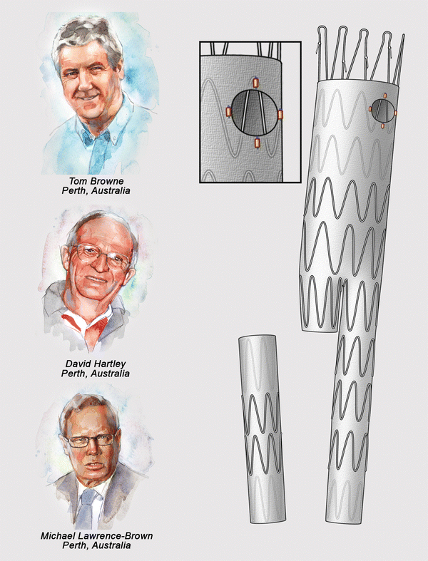
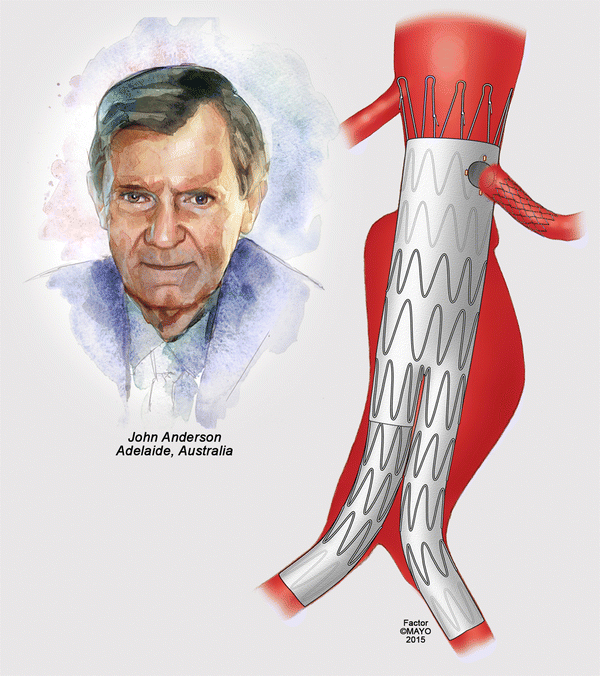
The first fenestrated repair was reported by Park in 1996 using a device modification to incorporate an accessory renal artery in a patient with infrarenal aneurysm. In 1997, Dr. Tom Browne (Fig. ). In the initial version of the device, two wires were used to constrain, and reduce the diameter of the device in its posterior wall. Today, fenestrated grafts with up to five fenestrations and scallops are routinely placed to treat short-neck AAA, juxtarenal AAA, pararenal AAA, and type IV TAAAs.
Fig. 1.2
The Perth research team of Tom Browne, David Hartley, and, led by, Michael Lawrence-Brown were instrumental in the initial animal experiments. The initial graft design was based on the Zenith Bifurcated Stent with suprarenal fixation. The fenestration was not reinforced and had radiopaque markers. Stent struts are noted across the fenestration, which at the time was not intended to be stented. The detailed designs and prototypes for the majority of the subsequent Cook production fenestrated and branched devices evolved out of the Perth R&D facility. By permission of Mayo Foundation for Medical Education and Research. All rights reserved
Fig. 1.3
The first clinical implant of a fenestrated stent using the Cook Zenith platform was performed by Dr. John Anderson in Adelaide, Australia. This illustration based on the actual case depicts a single left renal fenestration. The patient had been previously treated for a high-grade left renal stenosis by placement of a bare metal stent, which was carefully deployed inside the vessel. Note that the fenestration was non-reinforced and was not aligned by stent. By permission of Mayo Foundation for Medical Education and Research. All rights reserved
Fig. 1.4
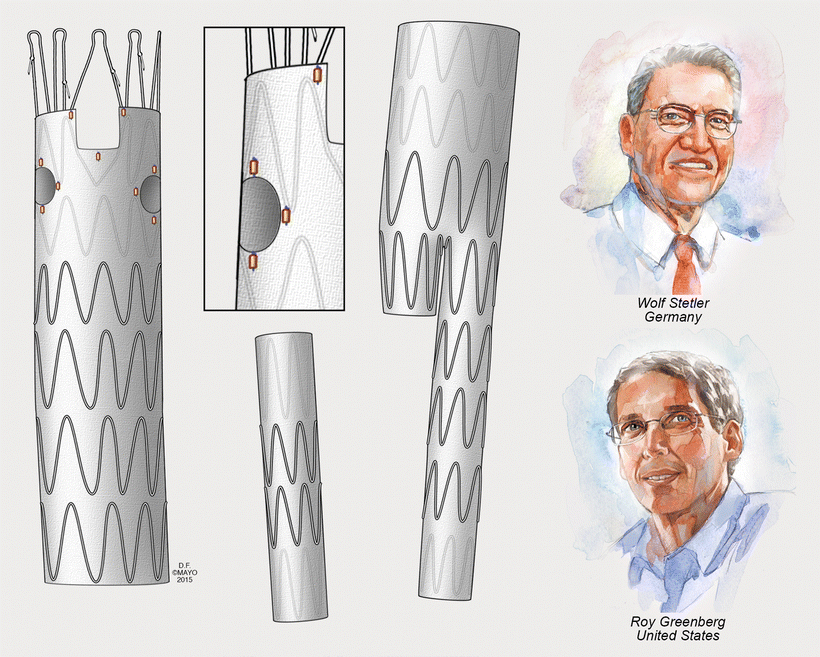
Progress in the design of the fenestrated graft is credited to Wolf Stelter in Germany who suggested the concept of a separate tubular component with the fenestrations and a distal bifurcated component. Roy Greenberg in the USA is credited with applying the technology to wide clinical use, treating complex anatomy with multiple fenestrations and scallops in a large number of patients. The fenestrations at this point were not reinforced, but the design had evolved to avoid struts across the fenestrations, with the intention to align the fenestration to the target vessel with an alignment stent. By permission of Mayo Foundation for Medical Education and Research. All rights reserved





![Shanks - Essential bicycle maintenance & repair: [step-by-step instructions to maintain and repair your road bike]](/uploads/posts/book/235248/thumbs/shanks-essential-bicycle-maintenance-repair.jpg)
