André Y. Denault Vegas Annette Yoan Lamarche - Basic Transesophageal and Critical Care Ultrasound
Here you can read online André Y. Denault Vegas Annette Yoan Lamarche - Basic Transesophageal and Critical Care Ultrasound full text of the book (entire story) in english for free. Download pdf and epub, get meaning, cover and reviews about this ebook. year: 2016, publisher: CRC Press, genre: Home and family. Description of the work, (preface) as well as reviews are available. Best literature library LitArk.com created for fans of good reading and offers a wide selection of genres:
Romance novel
Science fiction
Adventure
Detective
Science
History
Home and family
Prose
Art
Politics
Computer
Non-fiction
Religion
Business
Children
Humor
Choose a favorite category and find really read worthwhile books. Enjoy immersion in the world of imagination, feel the emotions of the characters or learn something new for yourself, make an fascinating discovery.
- Book:Basic Transesophageal and Critical Care Ultrasound
- Author:
- Publisher:CRC Press
- Genre:
- Year:2016
- Rating:4 / 5
- Favourites:Add to favourites
- Your mark:
Basic Transesophageal and Critical Care Ultrasound: summary, description and annotation
We offer to read an annotation, description, summary or preface (depends on what the author of the book "Basic Transesophageal and Critical Care Ultrasound" wrote himself). If you haven't found the necessary information about the book — write in the comments, we will try to find it.
André Y. Denault Vegas Annette Yoan Lamarche: author's other books
Who wrote Basic Transesophageal and Critical Care Ultrasound? Find out the surname, the name of the author of the book and a list of all author's works by series.


Basic Transesophageal and Critical Care Ultrasound — read online for free the complete book (whole text) full work
Below is the text of the book, divided by pages. System saving the place of the last page read, allows you to conveniently read the book "Basic Transesophageal and Critical Care Ultrasound" online for free, without having to search again every time where you left off. Put a bookmark, and you can go to the page where you finished reading at any time.
Font size:
Interval:
Bookmark:

Carl Chartrand-Lefebvre, Andr Y Denault and Annette Vegas
CT Correlation. antero posterior view, transverse plane view and sagittal plane view from computed tomographic images

https://youtu.be/T9U9pl-GKeE
https://youtu.be/va-HEt3M48g
https://youtu.be/ARaty0Ww-pQ
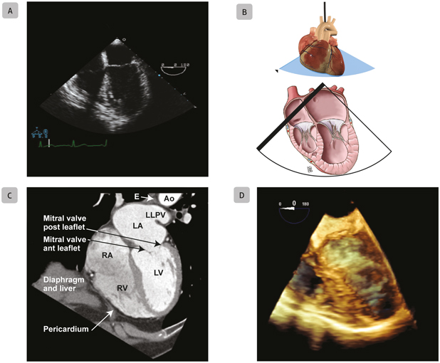
A1 Mid-Esophageal Four-Chamber. Mid-esophageal four-chamber views obtained with two-dimensional echocardiography ( A , B ), ECG-gated computed tomography ( C ), and three-dimensional echocardiography ( D ). Abbreviations: Ao, aorta; E, esophagus; ECG, electrocardiogram; LA, left atrium; LLPV, left lower pulmonary vein; LV, left ventricle; RA, right atrium; RV, right ventricle. Source: Illustration B courtesy of Gian-Marco Busato.
https://youtu.be/Ebj0jNtM2h8
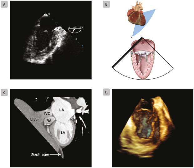
A2 Mid-Esophageal Two-Chamber Mitral Commissural. Mid-esophageal two-chamber mitral commissural views obtained with two-dimensional echocardiography ( A , B ), ECG-gated computed tomography ( C ), and three-dimensional echocardiography ( D ). Abbreviations: ECG, electrocardiogram; IVC, inferior vena cava; LA, left atrium; LV, left ventricle; RA, right atrium. Source: Illustration B courtesy of Gian-Marco Busato.
https://youtu.be/j2X-EC7Jkys

A3 Mid-Esophageal Two-Chamber. Mid-esophageal two-chamber views obtained with two-dimensional echocardiography ( A , B ), ECG-gated computed tomography ( C ), and three-dimensional echocardiography ( D ). Abbreviations: E, esophagus; ECG, electrocardiogram; LA, left atrium; LAA, left atrial appendage; LV, left ventricle. Source: Illustration B courtesy of Gian-Marco Busato.
https://youtu.be/bYotE-CB0TU
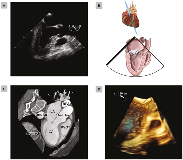
A4 Mid-Esophageal Long-Axis. Mid-esophageal long-axis views obtained with two-dimensional echocardiography ( A , B ), ECG-gated computed tomography ( C ), and three-dimensional echocardiography ( D ). Abbreviations: Ao, aorta; Asc, ascending; Des, descending; ECG, electrocardiogram; LA, left atrium; LV, left ventricle; RPA, right pulmonary artery; RVOT, right ventricular outflow tract. Source: Illustration B courtesy of Gian-Marco Busato.
https://youtu.be/EyvQmFyaSzY
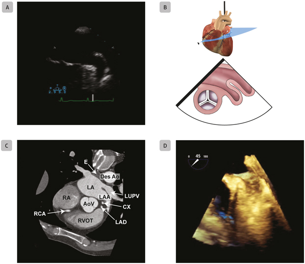
A5 Mid-Esophageal Left Atrial Appendage. Mid-esophageal left atrial appendage views obtained with two-dimensional echocardiography ( A , B ), ECG-gated computed tomography ( C ), and three-dimensional echocardiography ( D ). Abbreviations: Ao, aorta; AoV, aortic valve; CX, circumflex artery; Des, descending; E, esophagus; ECG, electrocardiogram; LA, left atrium; LAA, left atrial appandage; LAD, left anterior decending artery; LUPV, left upper pulmonary vein; RA, right atrium; RCA, right coronary artery; RVOT, right ventricular outflow tract. Source: Illustration B courtesy of Gian-Marco Busato.
A: https://youtu.be/Nt-EPvyiH6k
D: https://youtu.be/yQoGlkZA3Gc
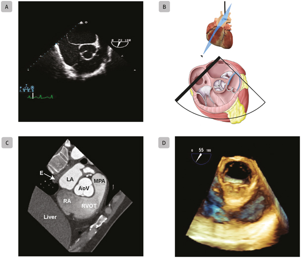
A6 Mid-Esophageal Right Ventricular Outflow Tract. Mid-esophageal right ventricular outflow tract views obtained with two-dimensional echocardiography ( A , B ), ECG-gated computed tomography ( C ), and three-dimensional echocardiography ( D ). Abbreviations: AoV, aortic valve; E, esophagus; ECG, electrocardiogram; LA, left atrium; MPA, main pulmonary artery; RA, right atrium; RVOT, right ventricular outflow tract. Source: Illustration B courtesy of Gian-Marco Busato.
A: https://youtu.be/x0fKqo71Aa8
D: https://youtu.be/nFXS_CHGOzM
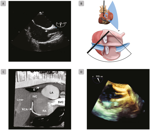
A7 Mid-Esophageal Bicaval. Mid-esophageal bicaval views obtained with two-dimensional echocardiography ( A , B ), ECG-gated computed tomography ( C ), and three-dimensional echocardiography ( D ). Abbreviations: E, esophagus; ECG, electrocardiogram; IVC, inferior vena cava; LA, left atrium; RA, right atrium; RCA, right coronary artery; RPA, right pulmonary artery; SVC superior vena cava. Source: Illustration B courtesy of Gian-Marco Busato.
https://youtu.be/qhywHNLzMG4
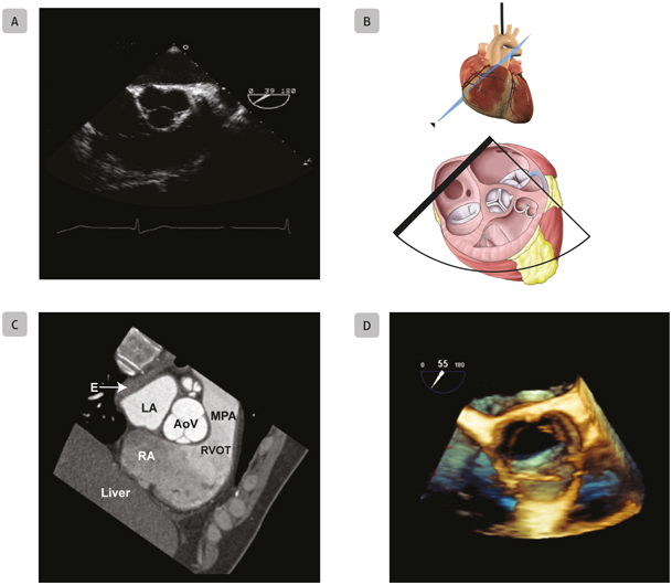
A8 Mid-Esophageal Aortic Valve Short-Axis. Mid-esophageal aortic valve short-axis views obtained with two-dimensional echocardiography ( A , B ), ECG-gated computed tomography ( C ), and three-dimensional echocardiography ( D ). Abbreviations: AoV, aortic valve; E, esophagus; ECG, electrocardiogram; LA, left atrium; MPA, main pulmonary artery; RA, right atrium; RVOT, right ventricular outflow tract. Source: Illustration B courtesy of Gian-Marco Busato.
A: https://youtu.be/_2R1RpJLAO8
Font size:
Interval:
Bookmark:
Similar books «Basic Transesophageal and Critical Care Ultrasound»
Look at similar books to Basic Transesophageal and Critical Care Ultrasound. We have selected literature similar in name and meaning in the hope of providing readers with more options to find new, interesting, not yet read works.
Discussion, reviews of the book Basic Transesophageal and Critical Care Ultrasound and just readers' own opinions. Leave your comments, write what you think about the work, its meaning or the main characters. Specify what exactly you liked and what you didn't like, and why you think so.