Some of the material in the fourth edition is derived from chapters in the third edition.
The editors acknowledge the contributions of the following authors to the previous edition.
William J. Frable
Catherine M. Keebler
Joseph F. Nasuti
Moira D. Wood
Maureen F. Zakowski
C. Meg McLachlin
Dr. Alan Nelson, VisionGate, Phoenix, AZ
Scott Mordecai and Dr. Frederic Preffer, Massachusetts General Hospital Pathology Department, Boston, MA
Basic Structure and Function of Mammalian Cells
Overview
Cells are the basic structural and functional units of all living organisms. Estimates about the total cell count of a human body vary widely, a number as large as 1014 has been proposed. Although the basic components of all cells are very similar, the differentiation of cells results in a wide variation of cellular morphology and function.
The smallest human cells, by diameter, are spermatozoa with a size of ~3 m, followed by the anucleate erythrocytes (8 m). The largest cells are female oocytes that can be as large as 3540 m and are visible to the naked eye. Motor neurons are extremely long cells, with their axons reaching from the spine to the distal extremities (up to 1.4 m in length).
Most cells can only be functional in large united structures, such as organs or suborganic structures. Other cells, mainly of hemato- or lymphopoietic origin, are mobile and autonomous, although, in many cases, their functionality is dependent on interaction with other cells.
Cytopathology studies diseases on the cellular level. While in histopathology, cells are assessed in the spatial context, in virtually all cytological applications, they are removed from their spatial context and have to be assessed isolated or as cell sheets.
Apart from the loss of the spatial information, cells can have considerably altered morphology when taken out of the united structures. Cellcell contacts are important features that build the shape of a cell. Many structural elements within a cell are connected to proteins that are attached to other cells or the basal membrane. This has to be considered when the same cell types are compared in histological versus cytological assessments.
Another important difference between histology and cytology is that, in histology, tissue sections are assessed 2-dimensionally, while in most cytology applications complete cells are analyzed that still have some 3-dimensional features, although they might appear flat in the microscope.
In this section, an overview of the most important cellular structures and functions that are relevant for cytopathology are presented (). We have assembled the most important information on cellular structures by describing the regular function in brief, the relevance for cytopathology and the morphology in normal and abnormal cells. For more detailed information about cellular structures and functions, a cell biology textbook is recommended.
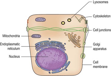
Figure 1-1 Schematic presentation of an epithelial cell displaying the most important structures discussed in this chapter.
Nucleus
The nucleus contains the genomic DNA, histones, and several proteins that are responsible for DNA replication, repair, and transcription of genetic information ().
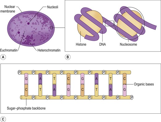
Figure 1-2 Contents of the nucleus, DNA . (A) A nucleus displaying nucleoli, euchromatin, and heterochromatin. (B) Two nucleosomes consisting of DNA coiled around histone proteins. (C) The structure of double stranded DNA. Organic bases are connected to a sugarphosphate backbone. Complementary bases (AT, CG) are held together by hydrogen bonds.
The assessment of a cell's nucleus is one of the most important tasks in cytopathology. The size of the normal nucleus is highly variable, depending on the underlying cell type. In many malignant cells, nuclei are considerably enlarged. Apart from nuclear size, the chromatin density, the nuclear membrane, and the presence of nucleoli are important features of nuclear morphology and will be described in detail.
The nucleus contains about 25% dry substance, of which 18% is DNA, plus a similar amount of histone proteins. The rest of the dry substance contains the non-histone proteins, nucleoli, and the nuclear membrane.
Contents of the Nucleus
DNA.
The genetic information of organisms is coded in deoxyribonucleic acid (DNA). DNA is a long stretch of single nucleotides that are connected by a sugarphosphate backbone (). The genetic information is stored in specific sequences consisting of four different bases: adenine, guanine, thymine, and cytosine (A, G, T, C). A triplet of bases is coding for an amino acid that constitutes the basic component of proteins. Although in principle the triplet code allows for 64 different variations, only 20 protein-building amino acids exist. Many amino acids are coded by multiple base triplets. The genetic code is degenerate, thereby tolerating errors in the base sequence to some degree. Two DNA stretches are combined as a double helix, one complete turn is reached after 10 bases. The DNA stretches are not covalently bound, but attached via hydrogen bonds between complementary bases AT and CG.
DNA is a very robust and stable molecule, since it has to protect the genetic code of an organism. The genetic information is transferred to the ribosomes (the protein production machinery) by ribonucleic acid (RNA), which has three important features different from DNA: RNA is based on a ribose backbone, it contains uracil instead of thymidine, and it is usually single-stranded. Compared with DNA, RNA is a rather unstable molecule.


