Springer-Verlag Berlin Heidelberg 2016
Stilianos E. Kountakis , Brent A. Senior and Wolfgang Draf (eds.) The Frontal Sinus 10.1007/978-3-662-48523-1_1
1. The Evolution of Frontal Sinus Surgery from Antiquity to the 21st Century
Adil A. Fatakia 1, Alla Y. Solyar 2 and Donald C. Lanza 2
(1)
West Jefferson Otolaryngology, New Orleans, LA, USA
(2)
Sinus & Nasal Institute of Florida Foundation, 550 94th Avenue North, St. Petersburg, FL 33702, USA
Donald C. Lanza
Email:
URL: http://www.sniflmd.com
Core Messages
Over the past 140 years, a rapid progression in the advancements of visualization and instrumentation has allowed for an evolution from open to endonasal techniques for the treatment of frontal sinus pathology.
Currently, endoscopic endonasal procedures have supplanted many open approaches given the low morbidity and comparable outcomes, but some advanced cases may require a combination of open and endonasal techniques as well as solely open approaches.
One lesson history has taught us is that re-establishing the natural drainage pathway of the frontal sinus into the ethmoid is a critical step in the management of most medically recalcitrant frontal sinus inflammatory disease
Introduction
Surgery of the frontal bone has existed for many millennia [].
Antiquity 1760 CE
Paleontologists and archeologists have demonstrated that otolaryngology, as well as neurosurgery, have their roots in what is believed amongst the earliest surgical procedure known to man called trepanation or trephination []. Opium, cocaine (Peru), and alcohol are among the earlier anesthetics available to aid in performing this procedure.
Examples of trepanation not only span time through to the present day but also span the globe [].
Fig. 1.1
Bronze age skull circa 3500 BCE with subacute osteomyelitis of the right maxillary and frontal sinus. Materials from excavation of burial ground Lchashen, (burial 52, 3035 years old). Consistent with the later description of Potts puffy tumor in 1760 CE []
Procedures involving the frontal sinuses per se were not formally described until long after they were first anatomically illustrated by Leonardo da Vinci in 1489 CE [].
Frontal Sinus Surgery 1750: Present
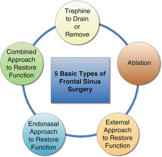
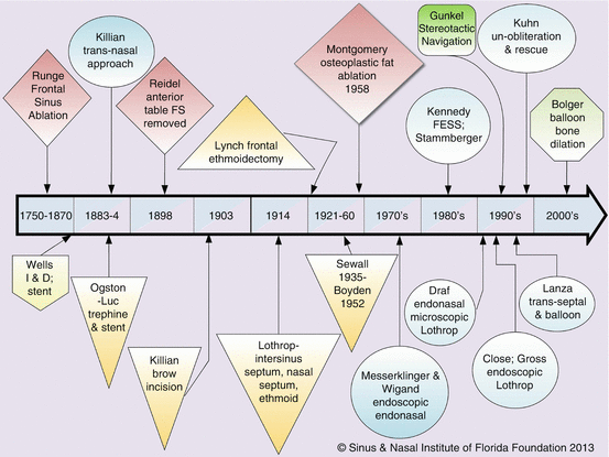
Over the last 140 years many procedures have been described to manage the unique challenges associated with an individualized patient approach and available technologies. The historical description that follows is divided into the varied surgical approaches that have shaped our current day frontal sinus surgery. These are: trephination, ablation, external approaches to restore function, endonasal approaches, endonasal balloon dilation, and sometimes combinations of external and endonasal approaches are applied to this day (See Figs. ).
Fig. 1.2
Historical types of frontal sinus surgery ( Permission granted by the Sinus & Nasal Institute of Florida Foundation 2013)
Fig. 1.3
History of frontal sinus timeline. Red diamonds = ablative procedures. Bone colored pentagon = trephination procedures. Blue ovals = endonasal approaches. Yellow triangles = external approaches with intention of restoring drainage. Grey-green octagon = balloon dilation without tissue removal. Green rectangle = technology introduction ( Permission granted by the Sinus & Nasal Institute of Florida Foundation 2013) (Color figure online)
Trephination and Drainage
As described earlier, trephination has been performed for many millennia. However, the first medical journal report of frontal sinus surgery appeared in the 1870 Lancet and described the work of Dr. Seolberg Wells in a man with a mucopyocele []. There was a high failure rate caused by frontal recess stenosis leading to abandonment of this procedure.
Ablation With and Without Reconstruction
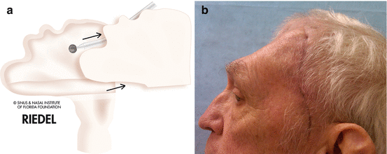
Ablation of the frontal sinus, whereby the mucosa is completely removed from within, is described from both an anterior and posterior table approach. The posterior table approach is typically performed during craniotomy for management of infection or malignancy. Although, Runge is said to have performed the first anterior frontal ablation in 1750 [].
Fig. 1.4
( a ) Schematic depiction of the Reidel procedure whereby the anterior table of the frontal is removed to gain access to ablate the frontal sinus ( Sinus & Nasal Institute of Florida Foundation 2013). ( b ) Later view of the deformity created by Reidel procedure employed in combination with neurosurgery for Postoperative infection in previously radiated patient with adenocarcinoma ( Permission granted by the Sinus & Nasal Institute of Florida Foundation 2013)
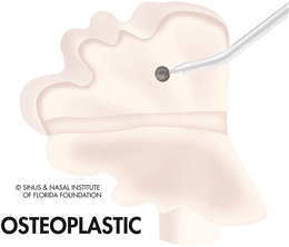
In an effort to minimize deformity, Hajek in 1903 proposed utilizing an osteoplastic flap whereby a hinged flap of anterior table frontal sinus bone was elevated with is periosteal blood supply attached [].
Fig. 1.5
Schematic depiction of the hinged osteoplastic flap with the sinus mucosa ablated from the lumen ( Permission granted by the Sinus & Nasal Institute of Florida Foundation 2013)
On the other hand, complications such as CSF leak and forehead paresthesia were seen more commonly. Additionally, delayed failures at 820 years with mucocele formation are not uncommon even today in the most experienced hands [].
External Fronto-Ethmoidectomy to Restore Drainage
In 1908, Dr. Knapp described performing extensive external ethmoidectomy through a medial orbital incision while enlarging the nasal frontal recess [).
Fig. 1.6
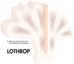
Schematic depiction of the Lothrop procedure indicating the removal of the frontal sinus intersinus septum, the nasal septum and creating one common opening to the paired frontal sinuses from medial orbit to orbit ( Permission granted by the Sinus & Nasal Institute of Florida Foundation 2013)


