Part I
Overview of Disorder
Springer International Publishing AG 2018
Pooya Khan Mohammad Beigi A Clinician's Guide to Pemphigus Vulgaris
1. Background
Pooya Khan Mohammad Beigi 1
(1)
Misdiagnosis Association & Society, Seattle, WA, USA
Keywords
Epidermal Basement membrane Anatomy Basal keratinocyte layer Lamina lucida Lamina densa Sublamina densa Epidermal pemphigus Subepidermal pemphigoid Pemphigus vulgaris Pemphigus foliaceus Paraneoplastic pemphigus
Basement membranes are thin layers of extracellular connective tissue that divide epithelial cells from the underlying connective tissue or from different types of cells []. Basement membranes are very complex structures that play different roles and have different compositions and structures, depending on the type of tissue they are found in. Functionally, basement membranes play many different roles. For instance, they are the site of attachment for many cells; can influence the behavior of cells such as their growth, apoptosis, development, and differentiation; and can also be the backbone structure for cell and tissue repair. The basement membrane can also regulate the extracellular environment of cells by acting as a selectively permeable structure. In this book, we will specifically be discussing the structure of the epidermal basement membrane, because many of the diseases that we will discuss will require a basic understanding of this important type of basement membrane.
The epidermal basement membrane consists of four major layers [).
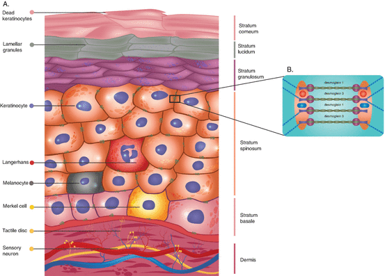
Fig. 1.1
Anatomy of the basement membrane . ( a ) The five layers of epidermis, which include the superficial layer of stratum corneum, stratum lucidum, stratum granulosum, stratum spinosum, and the innermost layer of stratum basale. Stratum corneum is made up of dead keratinocytes that are linked together with proteins. Stratum lucidum contains dead keratinocytes that have not completely finished the keratinization process. Stratum granulosum is the layer containing keratinocytes that contain keratohyalin granules. Stratum spinosum is characterized by the presence of desmosomes that results in an impermeable junction between keratinocytes as seen in section B of this image. Stratum basale is the deepest layer of epidermis, which contains a single layered column of epidermal stem cells. Moreover, the epidermis also contains specialized cells that are most prominent in stratum spinosum. For example, Langerhans are present in all stratums except stratum corneum, and their primary function is to fight skin infections by becoming antigen-presenting cells. Melanocytes are melanin-producing cells responsible for skin color. Merkel cells contain mechanoreceptors responsible for the detection of light touches. ( b ) The desmosome junction between keratinocytes that is created by the binding of desmoglein 1 (dsg1) and desmoglein 3 (dsg3) antibodies that attach to cell surface (Source: Pooya Khan Mohammad Beigi)
Acquired immune vesiculobullous diseases are diseases in which the body starts to produce autoantibodies against protein antigens in the epidermis or in the basement membrane associated with the epidermal cells []. Acquired immunobullous diseases can be divided into epidermal pemphigus or subepidermal pemphigoid diseases. Pemphigus diseases involve autoantibodies against proteins in the epidermis, whereas the pemphigoid group of diseases involves subepidermal autoantibodies against proteins in the basement membrane associated with the epidermis.
Pemphigus
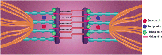
The pemphigus group of diseases are immune diseases involving immunoglobulin G (IgG) autoantibodies against various proteins found on the surface of epidermal cells [).
Fig. 1.2
The junction between keratinocytes that is characterized by desmosomes , which create an impermeable junction between the cells. The desmosome junction is created by the binding of desmoglein 1 (dsg1) and desmoglein 3 (dsg3) antibodies that attach to cell surface. As seen in the image, the junction is created with the help of additional proteins. For example, envoplakins interact with periplakins in order to serve as an intermediate filament in the desmosome junction. The plakoglobin serves as the cytoplasmic component. Lastly, plakophilins are proteins that link cadherins to intermediate filaments in the junction (Source: Pooya Khan Mohammad Beigi MD)
Desmoglein Compensation Hypothesis of Pemphigus Disease Presentation
On mucosal surfaces, both desmoglein 1 and 3 are expressed; however, desmoglein 3 dominates in terms of its expression in the mucosa compared to desmoglein 1 [].
Pemphigus vulgaris has two primary clinical variants, and the clinical features of these two variants can also be explained based on desmoglein expression. The mucocutaneous variant that involves autoantibodies against desmoglein 1 and 3 causes mucosal erosions and deep skin blisters [].
Types of Pemphigus Diseases
Pemphigus Vulgaris (Explained in Detail in the Next Section)
Pemphigus Vegetans
Pemphigus vegetans is a clinical variant of pemphigus vulgaris that involves erosions that form fungoid or papillomatous growths. These fungoid or vegetative growths are mainly seen on the scalp or face. This clinical variant is fairly rare and involves two subtypes: mild Hallopeau and severe Neumann types [].
Pemphigus Foliaceus
Pemphigus foliaceus only has cutaneous erosions and does not involve any mucosal erosions. It is an autoimmune disease in which the body produces IgG autoantibodies against desmoglein 1 proteins on the surface of keratinocytes in the superficial upper layers of the epidermis. The erosions look crusted and scaly in appearance and are transient in nature. These erosions also have an erythematous base. These erosions are found mainly on the upper trunk, scalp, and face [].
Pemphigus foliaceus is similar to pemphigus vulgaris with respect to the fragile blisters that are rarely ever found intact; thus, we only see the crust and scaly erosions left over from the ruptured superficial vesicles []. In this disease, the Nikolsky sign is present. The Nikolsky sign involves pushing the top of a blister and observing whether the top epidermal layers of the blister move laterally. This would indicate the absence of intercellular connections holding the top layers of epidermal cells together. Patients that have this disease often complain of painful burning lesions.
Pemphigus Erythematosus
This is also known as the Senear-Usher syndrome []. This is considered to be a type of pemphigus foliaceus that is only found on the malar region of the face. So it is a variant of pemphigus foliaceus, in which the crusted lesions only appear on the nose and malar regions.
Paraneoplastic Pemphigus
Paraneoplastic pemphigus is a disease associated with the presence of benign or malignant neoplasms []. The most commonly associated neoplasms include non-Hodgkins lymphoma and chronic lymphocytic leukemia, which account for around 67% of all the paraneoplastic pemphigus cases. Castlemans disease is associated with about 10% of the paraneoplastic pemphigus cases. This disease is not associated with common tumors, such as breast or colon carcinomas.


