Herbert B. Allen Dermatology Terminology 10.1007/978-1-84882-840-7_1 Springer-Verlag London Limited 2010
1. Papulosquamous Diseases
Abstract
Papulosquamous diseases are characterized by scaling papules and plaques. Scaling is synonymous with squamous in this regard. This group of diseases traditionally includes psoriasis, parapsoriasis, pityriasis rosea, pityriasis rubra pilaris, lichen planus, tinea corporis, secondary syphilis, mycosis fungoides, and drug eruptions. These diseases represent diverse infectious, proliferative, reactive, and neoplastic conditions.
The papules and plaques may have different shapes and sizes. Papules may be pinpoint, follicular, round, oval, polygonal, or even atrophic but are never square or rectangular. It is a truism of dermatology that nature abhors straight lines. Lesions with straight lines suggest extrinsic sources. Papules may be dome-shaped, acuminate (or pointed), ulcerated, verrucoid, telangiectatic, bleeding, oozing, excoriated, or crusted.
Papulosquamous diseases are characterized by scaling papules and plaques. Scaling is synonymous with squamous in this regard. This group of diseases traditionally includes psoriasis, parapsoriasis, pityriasis rosea, pityriasis rubra pilaris, lichen planus, tinea corporis, secondary syphilis, mycosis fungoides, and drug eruptions. These diseases represent diverse infectious, proliferative, reactive, and neoplastic conditions.
Figure 1.1.
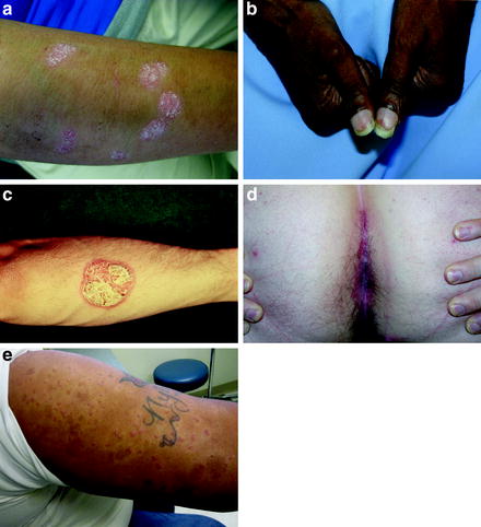
( a ) Silvery scaling is present on the plaques in psoriasis. ( b ) Yellow to red discoloration beneath the nails represents oil spots appearing like drops of oil on a piece of paper. ( c ) Auspitz sign is bleeding when scale is removed from a plaque. Hemorrhagic crusts represent this. ( d ) Redness and maceration in the natal cleft form the Abramowitz sign. ( e ) Woronoff rings are white rings around the psoriatic plaques.
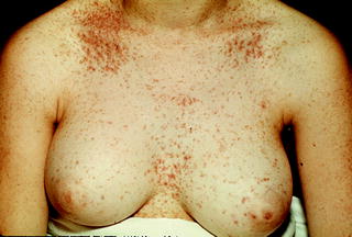
Figure 1.2.
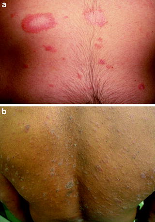
( a ) The herald patch is the largest lesion seen in pityriasis rosea. It predicts the more extensive eruption. ( b ) Christmas tree distribution refers to lesions that align on the trunk like the branches of an evergreen.
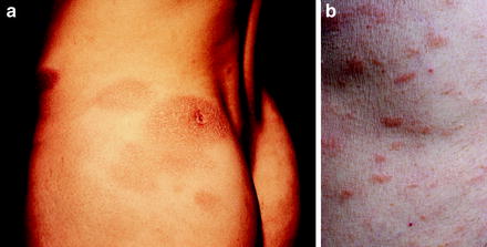
Figure 1.3.
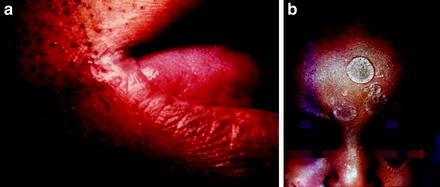
( a ) A split papule is a fissured papule at the angle of the mouth in secondary syphilis. ( b ) Small and medium-sized circular, scaling plaques represent nickel and dime lesions.
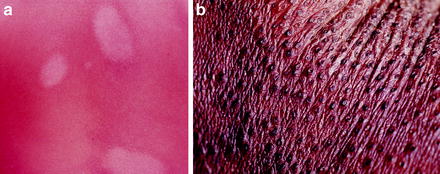
Figure 1.4.
( a ) Lesions of lichen planus are purple, pruritic, polygonal, scaling papules. ( b ) Wickham striae are white streaks on the purple plaques.
Figure 1.5.
Papules with a greasy feel are noted in Darier disease.
Figure 1.6.
( a) Fawn-colored patches are noted in parapsoriasis. ( b ) Oval digitate papules and plaques are seen in parapsoriasis.
Figure 1.7.
( a ) Islands of sparing in a confluent, red, scaling eruption are seen in pityriasis rubra pilaris. ( b ) Small, aggregated circular, horn-like papules make up the nutmeg grater.
The papules and plaques may have different shapes and sizes. Papules may be pinpoint, follicular, round, oval, polygonal, or even atrophic but are never square or rectangular. It is a truism of dermatology that nature abhors straight lines. Lesions with straight lines suggest extrinsic sources. Papules may be dome-shaped, acuminate (or pointed), ulcerated, verrucoid, telangiectatic, bleeding, oozing, excoriated, or crusted.
Colors of the papules range from white to black and almost every color in between, including multiple shades of red. The intensity of red often indicates chronicity in a lesion: i.e., the brighter the red, the more acute the lesion. A duller red generally indicates a resolving lesion. Red-pink, red-yellow, and red-orange often represent acute lesions, whereas red-blue, red-brown, and violet represent older lesions. Color recognition is very important, and individual colors make up some of the keywords, including blue for blue nevus, cherry red for carbon monoxide poisoning, and black for melanoma.
When scaling is identified together with papules, the spectrum of diseases narrows considerably and includes those diseases considered above. This classification is not perfect, however, and contains many exceptions. One of these is chronic atopic dermatitis, which may present with scaling papules or plaques but by convention is included in the dermatitis/eczema group of diseases. The chronic, acute, and subacute types of atopic dermatitis histologically have variable amounts of fluid in the epidermis (spongiosis), so this is perhaps why this disease is not classified in the papulosquamous group. Seborrheic dermatitis and lichen simplex chronicus are rashes with red scaling plaques but are also included in the dermatitis/eczema group even though they may present with a psoriasiform appearance. This rash also shows spongiosis histologically, similar to atopic dermatitis. Actinic keratosis, another exception, often presents as a red scaling papule, but this lesion is precancerous and not part of a rash. Seborrheic keratosis is also a scaling papule, but it is a tumor and not a rash. Other diseases that present with scaling plaques and a rash include Darier disease, erythrokeratodermia variabilis, and Netherton syndrome; however, these diseases are considered genetic disorders.
Scale may be further classified and may itself represent a keyword (e.g., micaceous in psoriasis and greasy in Darier disease). Further characterizations in scale also representing keywords include dirty for X-linked recessive ichthyosis; platelike for ichthyosis vulgaris; peripheral for pityriasis rosea; and trailing for erythema annulare centrifugum. Scale may be thin, and the thinness may be further classified, as in branny or cigarette paper as seen in pityriasis rosea. Scale may be thick or adherent, pinpoint or ostraceous, rocklike or nutmeg graterlike, or yellow or brown.
Carpet-tack scale is seen in discoid lupus erythematosus: when the scale is examined from underneath, spicules are seen to correspond to follicular orifices. Some authors consider discoid lupus erythematosus one of the papulosquamous diseases. I have included it here in the Dermatology in Systemic Disease chapter. Similarly, subacute cutaneous lupus erythematosus can be considered a papulosquamous disease because it often presents with plaques that have the appearance of psoriasis.


