Springer-Verlag London 2015
Herbert B. Allen The Etiology of Atopic Dermatitis 10.1007/978-1-4471-6545-3_1
1. Clinical Presentations
Herbert B. Allen 1
(1)
Department of Dermatology, Drexel University, Philadelphia, Pennsylvania, USA
Abstract
This chapter presents a pictorial representation of the variations of atopic dermatitis featured in this work, including facial-extensor, flexural, and nummular. Less frequently occurring variants are also represented, including lichen planuslike eczema, dyshidrotic eczema, pityriasis alba, and juvenile plantar dermatosis. Images also include diseases in which eczema is a known component (Meyersons nevus and Doucas Kapetanakis pigmented purpuric dermatosis) and diseases in which eczema was previously not thought to be present (seborrheic dermatitis, tinea pedis, and axillary granular parakeratosis).
Keywords
Atopic dermatitis Axillary granular parakeratosis Doucas Kapetanakis pigmented purpuric dermatosis Dyshidrotic eczema Facial-extensor eczema Flexural eczema Juvenile plantar dermatosis Lichen planuslike eczema Meyersons nevus Nummular eczema Pityriasis alba Seborrheic dermatitis Tinea pedis
This chapter presents a pictorial representation of the variations of atopic dermatitis that are featured in the body of this work. Heavily represented are the most common subtypes of atopic dermatitis: facial-extensor (Figs. ), are also shown.
Each of these disorders may overlap with others, so many times they are not pure forms of the variant. This happens relatively frequently in the facial-extensor variety presenting with flexural involvement, or with facial extensor concomitantly occurring with nummular disease. Pityriasis alba occurs very frequently with all of the major forms as well as occurring in a solo fashion. Dyshidrotic eczema may occasionally overlap (in my observations, most often with flexural disease). Other variants are rare enough that overlap with other forms of the disease is very uncommon. In addition to overlap, each of these may become impetiginized, with certain exceptions, such as pityriasis alba. The images are mostly from our patients at Drexel Dermatology; some have been graciously supplied by the Wake Forest Department of Dermatology.
Facial
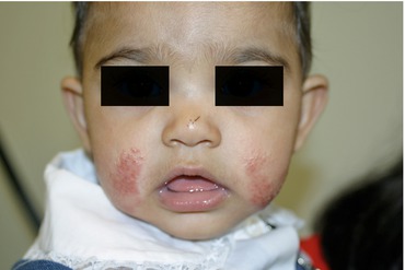
Fig. 1.1
Atopic dermatitis, facial-extensor. In this infant, there are crusted, pink-red papulovesicular plaques on bilateral cheeks
Fig. 1.2
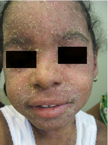
Atopic dermatitis, facial-extensor. Marked yellowish scaling covering the entire face, and underlying pink edematous plaques, are present in this child
Fig. 1.3
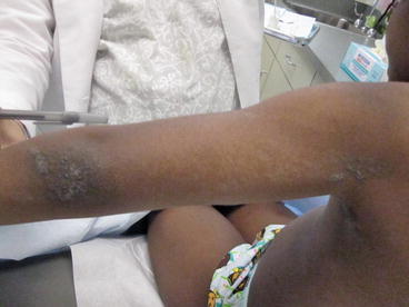
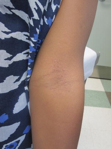
Atopic dermatitis, facial-extensor (arm). On the arm is a dull red, hyperpigmented papular eruption; small lichenified plaques are also present
Fig. 1.4
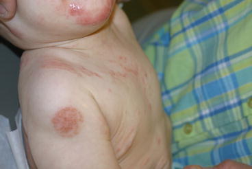
Atopic dermatitis, facial-extensor nummular overlap. On the chin of this infant are red weeping papulovesicular plaques; on the shoulder is a circular, dull pink, slightly crusted plaque
Fig. 1.5
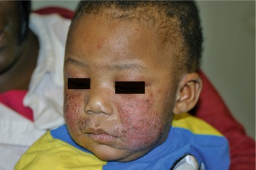
Atopic dermatitis, facial-extensor impetiginized. Crusted, edematous, pink plaques are present on bilateral cheeks and the upper lip in this toddler
Flexural
Fig. 1.6
Atopic dermatitis, flexural (mild). In this childs antecubital fossa is an eruption of faint pink papulovesicles. (This may be the first visible change in this disease; the flesh-colored papules are likely the primary lesions)
Fig. 1.7
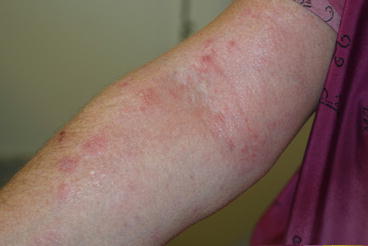
Atopic dermatitis, flexural (severe). On this antecubital fossa, there is a lichenified, pink, scaling, crusted plaque with adjacent pink, scaling, crusted papules and plaques
Fig. 1.8
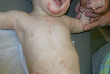
Atopic dermatitis, flexural facial extensor and nummular overlap. On this infants face, flexures, and trunk are red and pink plaques. The facial lesions are more severe
Fig. 1.9
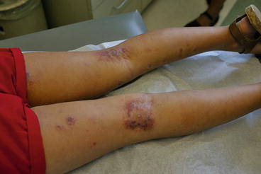
Atopic dermatitis, flexural (severe and impetiginized). On the bilateral popliteal fossae are dull red, eroded, crusted thickened plaques. Crusted edematous plaques are scattered on the rest of the legs
Nummular
Fig. 1.10
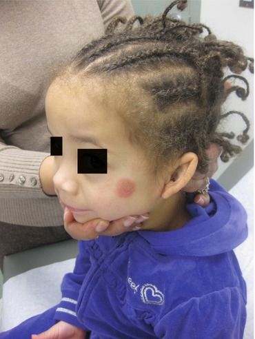
Atopic dermatitis, nummular (mild). On this childs face is a circular, red edematous plaque
Fig. 1.11
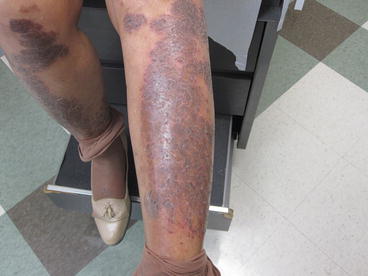
Atopic dermatitis, nummular (more severe). Large circular and confluent dull red-brown plaques cover bilateral shins in this woman
Fig. 1.12
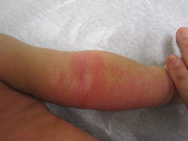
Atopic dermatitis, nummular (generalized). On this childs arm and trunk are bright red, confluent patches and plaques
Fig. 1.13
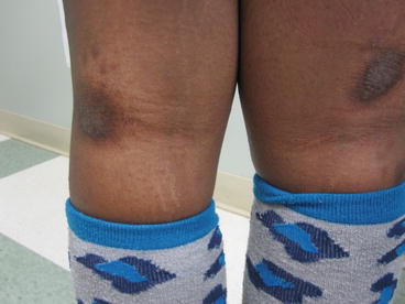
Atopic dermatitis, nummular, flexural overlap. On the left popliteal fossa is a brown, scale-crusted lichenified, oval plaque; superior to the right popliteal fossa is a similar plaque
Lichen PlanusLike
Fig. 1.14
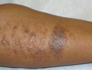
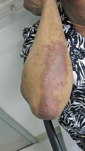
Lichen planuslike atopic dermatitis, local. Hyperpigmented, pink-brown papules and plaques are present on the medial forearm. Lichenification and scaling are also present focally

