Part 1
Anatomy, Pathology, and Pathophysiology
Charles Eaton , M. Heinrich Seegenschmiedt , Ardeshir Bayat , Giulio Gabbiani , Paul Werker and Wolfgang Wach (eds.) Dupuytrens Disease and Related Hyperproliferative Disorders 2012 Principles, Research, and Clinical Perspectives 10.1007/978-3-642-22697-7_1 Springer-Verlag Berlin Heidelberg 2012
1. Dupuytrens Disease: Anatomy, Pathology, and Presentation
Ghazi M. Rayan 1
(1)
Baptist Medical Centre, University of Oklahoma, Oklahoma City, OK, USA
1.1
1.1.1
1.1.2
1.1.3
1.1.4
1.1.5
1.2
1.2.1
1.2.2
1.2.3
1.2.4
1.2.5
1.3
1.3.1
1.3.2
1.3.3
1.3.4
1.3.5
1.4
1.4.1
1.4.2
Abstract
Dupuytrens disease has been recognized for approximately four centuries. A thorough knowledge of the palmar fascial complex anatomy is imperative for understanding Dupuytren disease pathology. Its presentation, although seemingly constant, is actually variable, depending on the structures involved. Dorsal skin changes in Dupuytren disease are a source of confusion. These can be either in the form of dorsal Dupuytrens nodules, which are similar to palmar nodules and pathognomonic of the disease or dorsal cutaneous pads, which are encountered equally in Dupuytren disease and in the normal population. There are also two distinct clinical entities associated with palmar fascial proliferation, classic Dupuytren disease and atypical, so-called non-Dupuytren disease. These two types differ in presentation, etiology, treatment, and prognosis. Authors of future epidemiological and outcome studies should not confuse these two clinical entities.
Keywords
Anatomy Pathology Non-Dupuytrens disease Dorsal Dupuytrens nodules Dorsal cutaneous pads Nodules (dorsal)
1.1 Anatomy
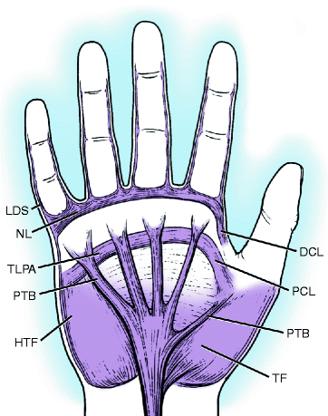
The palmar fascial complex (PFC) of the hand has five components (Rayan ).
Fig. 1.1
Illustration of the palmar fascial complex. DCL distal commissural ligament, HTF hypothenar muscle fascia, LDS lateral digital sheet, NL natatory ligament, PCL proximal commissural ligament, PTB pretendinous band (small finger and thumb bands tagged), TF thenar fascia, TLPA transverse ligament of the palmar aponeurosis. (From Rayan )
1.1.1 Radial Aponeurosis
Tubiana et al. () revealed that the thenar fascia is an extension of the CA and the pretendinous band is less defined than all other bands.
1.1.2 Ulnar Aponeurosis
This consists of the hypothenar muscle fascia, pretendinous band to the small finger and abductor digiti minimi (ADM) coalescence (White ) revealed that the hypothenar fascia is an extension of the CA and the pretendinous band is always present and very substantial.
1.1.3 Central Aponeurosis
The CA is the core of Dupuytrens disease (DD) activity. It is a triangular fascial layer with proximal apex and distal base and its fibers are oriented into three dimensions: longitudinal, transverse, and vertical.
(a)
Longitudinal fibers of the CA fan out as three pretendinous bands (PB) for the central digits and each bifurcate distally. McGrouther () described three microscopic layers of insertions for the split PB. The superficial layer inserts into the dermis, the middle continues to the digit as the spiral band, and the deep layer passes almost vertically in the vicinity of the extensor tendon.
(b)
Transverse Fibers of the CA encompass the natatory ligament (NL) which is distally located in the palmodigital region and the transverse ligament of the palmar aponeurosis (TLPA). The TLPA is located proximal and parallel to the NL and deep to the pretendinous bands. This ligament continues radially as the proximal commissural ligament. The TLPA gives origin to the septa of Legueu and Juvara (SLJ), protects the neurovascular (NV) structures, and provides an additional pulley to the flexor tendons.
(c)
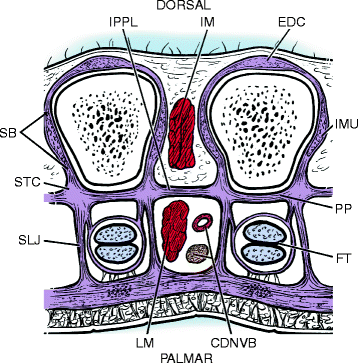
Vertical fibers of CA encompass the vertical bands of Grapow and the SLJ (Fig. ).
Fig. 1.2
Illustration of the interpalmar plate ligaments, septa of Legueu and Juvara, and soft tissue confluence. CDNVB Common digital neurovascular bundle, EDC extensor digitorum communis, FT flexor tendons, IM interosseous muscles, IMU intrinsic musculotendinous unit, IPPL interpalmar plate ligament, LM lumbrical muscle, PP palmar plate, SB sagittal band, SLJ septum of Legueu and Juvara, STC soft tissue confluence. (From Rayan )
Grapow () identified eight septa, one radial and ulnar for each digit. The radial were longer than the ulnar septa. The septa attach to the TLPA superficially and to a soft tissue confluence deeply and distally and to the interosseous muscle fascia proximally. The soft-tissue confluence consists of the SLJ, the interpalmar plate ligaments (IPPL), palmar plate, annular pulley, and sagittal band. Three IPPLs, radial, central, and ulnar form the floor (i.e., dorsal) of the three NV web space canals.
The septa form seven compartments of two types; four flexor septal canals that contained the flexor tendons, and three web space canals that contained common digital nerves and arteries and lumbrical muscles. Grossly and histologically, the septa are thicker and consisted of organized collagen distally but not proximally.
1.1.4 Palmodigital Fascia
Gosset () described several fascial structures that coalesce in the web-digital area, the most notable of these are the spiral band and NL. Each spiral band begins proximally as the middle layer of a bifurcated pretendinous band, tangential to the palm, then spirals on its axis, becoming perpendicular to the palm distally, lateral to the MP joint capsule. They course distally deep to the NV bundle and NL, and emerge distal to the NL to continue as the lateral digital sheet (LDS). The spiral band therefore is the connection between the palmar and digital fascial structures. The fibers of the NL run in a transverse plane, but the distal most fibers form a U and run longitudinally along the side of adjacent digits toward the LDS. The fibers of the NL run from side to side at the web spaces and extend radially to the first web space as the distal commissural ligament. The NL has a well-defined proximal border that becomes more pronounced when the fingers are fully abducted. The LDS therefore has deep and superficial contributions from the spiral band and NL.
1.1.5 Digital Fascia
The NV bundle in the digit is surrounded by four fascial structures; Graysons ligament palmar, Clelands ligaments dorsally, Gosset LDS laterally and possibly a retrovascular fascia (Thomine ) medially and dorsally that has not been confirmed by other studies. Some doubt the existence of the retrovascular fascia.
1.2 Pathology
Luck () considered the diseased cords and nodules to be the consequence of pathologic changes in the normal fascia. Dupuytrens nodules and cords are pathognomonic of DD. A nodule often precedes the cord, but sometimes a cord develops without a nodule. The cords typically shorten progressively leading to joint and tissue contracture. The cords are located in the palmar, palmodigital, or digital regions. Joint deformity in early stages is caused by diseased cords whereas long-standing flexion deformity can lead to capsuloligamentous tissue scarring with resultant joint contracture.

