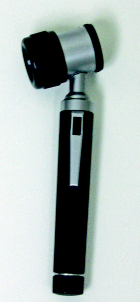
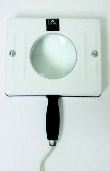
The dermatological exam begins with physicians questions to the patients about their skin condition and related symptoms. Demographic data, including age, gender, and race, besides previous conditions, medications, and family medical history are important elements. The skin should be always evaluated in a well-illuminated place, with direct light. The exam is performed from head to toe, including hair, mucosa, nails, and ganglions. It is also recommended using instruments, such as dermatoscope, Woods lamp, and digital photography, according to each patients needs.
1.2.1.3 Photographic Documentation
Photographic assessment of the skin can be important to record patients medical history, to follow up patients, and also when a second opinion is sought. Photographic assessment significantly improves patients understanding on their diagnostic and treatment progression [].
Before acquiring the images, it is suggested to ask patients to sign an informed consent form for photographs, especially if the patient can be recognized. Define high-quality standards to create and maintain photographic patient records as well as to guarantee and maintain patient anonymity and confidentially [].
Standard photographic methodology is recommended to collect and store patients images. The images should be always taken using the same parameters, such as camera settings, patient position, and light. Some pictures require a point-source flash, while others require elimination of shadows caused by using a ring flash [].
The minimum setup needed to document face and body is composed by digital camera; proper light source; appropriate computer to store, analyze, and display the digital files; and a trained photographer.
The photographer is responsible for controlling the standards previously defined when taking the photographs. Moreover, he/she must be patient, especially early on to keep the patient calm to achieve good quality images.
Most of the current digital cameras available in the market offer high resolution. For dermatological use, a resolution of four million pixels is enough []. Low-resolution images should be avoided.
Light source positioning is crucial for the photograph quality. Wrong positioning of the lights can create shadows, compromising the skin evaluation. The background must be neutral, monochromatic, and non-reflective, preferably dark. A dark and opaque background provides greater control of the illumination over the subject. The positioning of light source should be the same at all time points for the same subject.
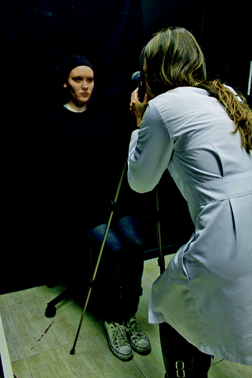
The relatively equal position of the subject to the camera enables the acquisition of the same field size before and after treatment. Makeup, jewelry, and clothing that might interfere the images should be removed. For facial photographs, usual positions are front, oblique view (45), and lateral (left and right), and a neutral face expression during the shoot is required (Fig. ).
Fig. 1.3
Photographic studio
A standardized setup and imaging procedure is recommended for better correlations between before and after treatment images. Further correlation between the two images could be accomplished using anatomical landmarks. The after treatment image should be compared to the before image immediately after acquisition for consistency and to retake if necessary.
Some objective imaging tools were developed to standardize the photographic position and to assure the photographic quality. Companies like Canfield ScientificTM and FotoFinder SystemsTM developed a series of methods and equipments to obtain standardized, reproducible, serial medical photographs and documentation reducing the photographic variables of images.


