• Complain

David J. Browning - Hydroxychloroquine and Chloroquine Retinopathy

Here you can read online David J. Browning - Hydroxychloroquine and Chloroquine Retinopathy full text of the book (entire story) in english for free. Download pdf and epub, get meaning, cover and reviews about this ebook. year: 2014, publisher: Springer-Verlag New York Inc., genre: Romance novel. Description of the work, (preface) as well as reviews are available. Best literature library LitArk.com created for fans of good reading and offers a wide selection of genres:

Romance novel Science fiction Adventure Detective Science History Home and family Prose Art Politics Computer Non-fiction Religion Business Children Humor

Choose a favorite category and find really read worthwhile books. Enjoy immersion in the world of imagination, feel the emotions of the characters or learn something new for yourself, make an fascinating discovery.

David J. Browning Hydroxychloroquine and Chloroquine Retinopathy
  • Book:
    Hydroxychloroquine and Chloroquine Retinopathy
  • Author:
  • Publisher:
    Springer-Verlag New York Inc.
  • Genre:
  • Year:
    2014
  • Rating:
    3 / 5
  • Favourites:
    Add to favourites
  • Your mark:
    • 60
    • 1
    • 2
    • 3
    • 4
    • 5

Hydroxychloroquine and Chloroquine Retinopathy: summary, description and annotation

We offer to read an annotation, description, summary or preface (depends on what the author of the book "Hydroxychloroquine and Chloroquine Retinopathy" wrote himself). If you haven't found the necessary information about the book — write in the comments, we will try to find it.

This book covers the clinical management of ocular toxicity of hydroxychloroquine and chloroquine for the practicing ophthalmologist, rheumatologist and dermatologist. It also details practice management considerations as well as medico-legal responsibilities.

David J. Browning: author's other books


Who wrote Hydroxychloroquine and Chloroquine Retinopathy? Find out the surname, the name of the author of the book and a list of all author's works by series.

Hydroxychloroquine and Chloroquine Retinopathy — read online for free the complete book (whole text) full work

Below is the text of the book, divided by pages. System saving the place of the last page read, allows you to conveniently read the book "Hydroxychloroquine and Chloroquine Retinopathy" online for free, without having to search again every time where you left off. Put a bookmark, and you can go to the page where you finished reading at any time.

Light

Font size:

Reset

Interval:

Bookmark:

Make
David J. Browning Hydroxychloroquine and Chloroquine Retinopathy 2014 10.1007/978-1-4939-0597-3_1
Springer Science+Business Media New York 2014
1. Preclinical Foundations: Relevant Anatomy and Physiology
David J. Browning 1
(1)
Charlotte Eye Ear Nose & Throat Associates, Charlotte, NC, USA
Abstract
Reviewing the preclinical science relevant to the mechanisms and risk factors for chloroquine and hydroxychloroquine retinopathy can solidify clinical understanding of that condition. This chapter gathers the scattered and often difficult-to-access pertinent facts and concepts.
Abbreviations
4AQs
4-Aminoquinolines (chloroquine and hydroxychloroquine)
APC
Antigen presenting cell
4AQR
4-Aminoquinoline retinopathy
BM
Bruchs membrane
BRB
Bloodretina barrier
C
Concentration
CpG ODN
CpG oligodeoxynucleotide
CD
CD number
CD3
Cluster of differentiation T3 cell co-receptor
CD74 gene
Cluster of differentiation 74 gene
D
Daily dose in mg/kg
DN cells
Double negative cells
DP cells
Double positive cells
EC50
Effective concentration 50 %
ELM
External limiting membrane
FAZ
Foveal avascular zone
GCL
Ganglion cell layer
HLA
Human leukocyte antigen
IFN
Interferon alpha
Ii
Invariant chain
IK complex
Inhibitor of kappa B kinase complex
IL
Interleukin
ILM
Internal limiting membrane
INL
Inner nuclear layer
IPL
Inner plexiform layer
IRAK
Interleukin-1 receptor-associated kinase
IS/OS
Inner segment/outer segment
K
Rate constant for elimination
m
Micrometer
MHC
Major histocompatibility complex
miRNA
MicroRNA
nm
Nanometer
NEMO
Nuclear factor- essential modifier regulatory subunit
NF-
Nuclear factor-
NFL
Nerve fiber layer
NLRs
Nucleotide-binding and oligomerizing domain-like receptors
OCT
Optical coherence tomography
OPL
Outer plexiform layer
ONL
Outer nuclear layer
PAMP
Pathogen-associated molecular pattern
RA
Rheumatoid arthritis
RCS
Royal College of Surgeons
RPC
Radial peripapillary capillary
RPE
Retinal pigment epithelium
SD-OCT
Spectral domain optical coherence tomography
SLE
Systemic lupus erythematosus
SP cells
Single positive cells
TCR
T cell receptor
TCRCD3
T cell receptorcluster of differentiation 3T cell co-receptor complex
TD-OCT
Time domain optical coherence tomography
Th cells
Helper T cells
Treg cells
Regulatory T cells
V
Volume of distribution
VEGF
Vascular endothelial growth factor
Concepts and facts in this chapter are drawn from ocular anatomy, physiology, immunology, and pharmacology that are important in understanding the cause, progression, and ways to prevent chloroquine and hydroxychloroquine retinopathy. Because chloroquine and hydroxychloroquine are 4-aminoquinolines (4AQs), the retinopathy that they cause is referred to as 4-aminoquinoline retinopathy (4AQR). It is assumed that the reader has a medical-school background but needs to be refreshed about the relevant topics. Where possible, the particular structures and physiologic steps influenced by 4AQs will be noted. Looking ahead, Chap. their toxicology and the pathologic ocular changes they can cause. Commonly used abbreviations in this chapter are collected in Abbreviations for reference. Each term will be first used in its full form, along with its abbreviation.
1.1 Anatomy and Histology
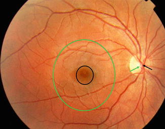
Histologically, the retina is a multilayered sheet of neuronal, glial, and vascular tissue that lines the inside posterior two-thirds of the eye. It is bounded anteriorly by the vitreous humor and posteriorly by the retinal pigment epithelium (RPE), Bruchs membrane (BM), and choroid. The macula is a circular area of the retina 5.25.5 mm in diameter with a center located 17, or 4.05.0 mm temporal, and 0.530.8 mm inferior to the center of the optic disc (Fig. ].
Fig 11 Fundus photograph from a normal eye showing the macula green circle - photo 1
Fig. 1.1
Fundus photograph from a normal eye showing the macula ( green circle ) and fovea ( black circle ). The normal central retinal artery ( black arrow ) is located nasal to the central retinal vein ( green arrow ) in the optic disc. The luteal pigment gives the region encircled in black its darker pigmentation relative to the surrounding retina
Xanthophylls are not synthesized by the body, but must come from the diet [].
The central 1.5 mm circular area of the macula is called the fovea, denoted by a gently curved depression in the retinal surface. Within the fovea is a roughly circular avascular area, the foveal avascular zone (FAZ), approximately 400500 m in diameter which contains only cones, present at a density of approximately 125,500140,000/mm2 [].
Deriving the Conversion from Degrees to Micrometers
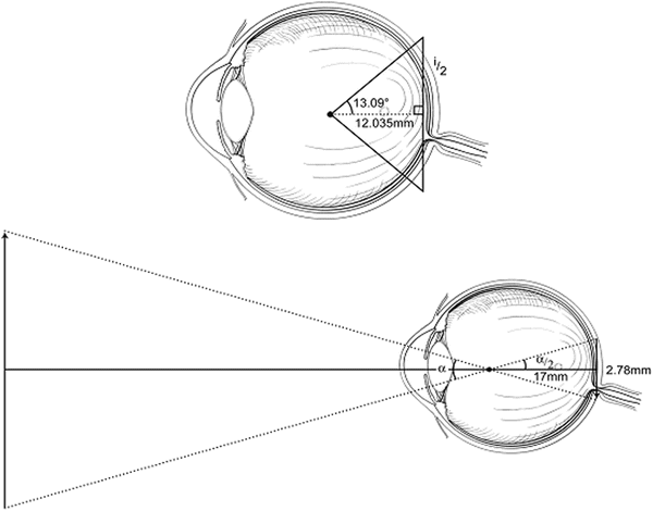
Without derivation, it has been asserted that 1280 m [):
Fig 12 Diagram of schematic eye used in deriving the conversion factor for - photo 2
Fig. 1.2
Diagram of schematic eye used in deriving the conversion factor for transforming arc length on the retina to subtended angle
360 (5.5/circumference of eye)=26.18.
One half the straightened image height ( i /2) can be calculated noting the geometrical relationship:
Solving for i and therefore i 2278 mm The simplified version of the eye - photo 3
Solving for i ,
and therefore i 2278 mm The simplified version of the eye is that of a - photo 4
and therefore i /2=2.78 mm.
The simplified version of the eye is that of a single lens 17 mm in front of the retina (the eyes nodal point). Ray tracing shows the relationship between object and image in Fig.. One half the angle subtended by the image from the eyes nodal point can be calculated from the geometrical relationship
Hydroxychloroquine and Chloroquine Retinopathy - image 5
where
The conclusion then is that 185755 mm or 10296 mm296 m which is in - photo 6
The conclusion, then, is that 18.57=5.5 mm, or 1=0.296 mm=296 m, which is in fair agreement with the published figure.
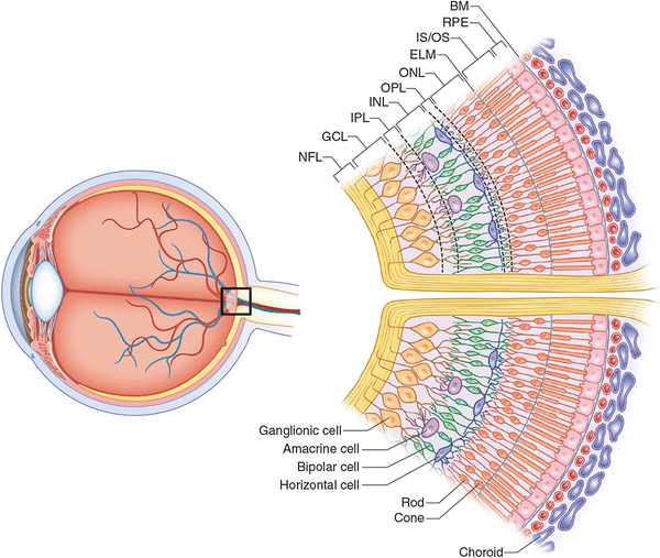
A cross section through the retina just outside the area centralis shows ten layers (Figs. ].
Fig 13 Diagram of the stratified cellular nature of the retina The axons of - photo 7
Next page
Light

Font size:

Reset

Interval:

Bookmark:

Make

Similar books «Hydroxychloroquine and Chloroquine Retinopathy»

Look at similar books to Hydroxychloroquine and Chloroquine Retinopathy. We have selected literature similar in name and meaning in the hope of providing readers with more options to find new, interesting, not yet read works.


Reviews about «Hydroxychloroquine and Chloroquine Retinopathy»

Discussion, reviews of the book Hydroxychloroquine and Chloroquine Retinopathy and just readers' own opinions. Leave your comments, write what you think about the work, its meaning or the main characters. Specify what exactly you liked and what you didn't like, and why you think so.