ISBN 978-0957149946
eISBN 978-1-910399-07-1
Text, design and illustration Zeshan Qureshi 2014
Edited by Mark Rodrigues and Zeshan Qureshi
Published by Zeshan Qureshi. First published 2014
All rights reserved; no part of this publication may be reproduced, stored in a retrieval system, transmitted in any form, or by any means, electronic, mechanical, photocopying, recording, or otherwise, without the prior written permission of the publishers.
Original design by Zeshan Qureshi and Mark Rodrigues. Page make-up by Anchorprint Group Limited
A catalogue record for this book is available from the British Library.
Publisher and Editors Acknowledgements:
We would like to thank all the authors for their hard work, and our panel of student reviewers for their unique input. We are extremely grateful for the support given by medical schools across the UK. We would also like to thank the medical students that have inspired this project, believed in this project, and have helped contribute to, promote, and distribute the book across the UK.
We would like to thank Dr Alan Grundy (Consultant Radiologist, St Georges Hospital, London) for providing the X-ray used in of the chest X-ray chapter.
Commissioned medical illustrations by:
Peter Gardiner ()
Although we have tried to trace and contact copyright holders before publication, in some cases this may not have been possible. If contacted, we will be pleased to rectify any errors or omissions at the earliest opportunity. Knowledge and best practice in this field are constantly changing. As new research and experience broaden our understanding, changes in research methods, professional practices, or medical treatment may become necessary. Practitioners and researchers must always rely on their own experience and knowledge in evaluating and using any information, methods, compounds, or experiments described herein. In using such information or methods, they should be mindful of their own safety and the safety of others, including parties for whom they have a professional responsibility. With respect to any drug or pharmaceutical products identified, readers are advised to check the most current information provided (i) on procedures featured or (ii) by the manufacturer of each product to be administered, to verify the recommended dose or formula, the method and duration of administration, and contraindications. It is the responsibility of practitioners, relying on their own experience and knowledge of their patients, to make diagnoses, to determine dosages and the best treatment for each individual patient, and to take all appropriate safety precautions. To the fullest extent of the law, neither the Publisher nor the authors, contributors, or editors, assume any liability for any injury and/or damage to persons or property that may occur as a result of any person acting or not acting based on information contained in this book
Printed and bound by Cambrian Printers in UK
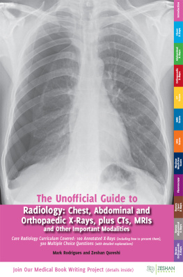
The Unofficial Guide to Radiology is the fifth book in the Unofficial Guide to Medicine series. Almost every patient has some form of medical imaging performed during his or her investigations and management. The commonest form of imaging, and the modality which all doctors should be able to interpret, remains the X-ray. This important aspect of radiology is therefore the main focus of the book. Other imaging modalities are specialised investigations interpreted by radiologists. However, medical students, doctors, nurses, physicians associates, and surgeons need to understand what these tests involve, when they are indicated and contraindicated, and how to best request them. Therefore, these aspects are also covered in the book.
Despite its universal importance, X-ray interpretation is often an overlooked subject in the medical school curriculum, which many medical students and junior doctors find difficult and daunting. I was no different when I started work as a junior doctor. However, since starting radiology training, I have realised X-ray interpretation should not be that way.
The keys to interpreting X-rays are having a systematic method for assessing the X-ray and getting lots of practice at looking at and presenting X-rays. Occasionally, there may be a complex X-ray you find difficult, or a subtle finding you overlook, but thats what keeps people like me in a job, so do not worry about it.
The 4 Ds are a useful framework for X-ray interpretation which underpins the approach used in this book. First, you need to Detect and Describe the abnormalities on the X-ray. You then need to form a Differential diagnosis based on clinical and X-ray findings before Deciding what further imaging and management is required.
There are lots of radiology textbooks available, but I do not think there is one which is ideally suited for teaching medical students and junior doctors. Many have small, often poor quality images. Radiology is a visual subject and therefore such images are difficult to use to demonstrate key clinical findings. This is confounded by the fact that the findings are usually only described in a figure below the image, and it is often difficult to know exactly what part of the image corresponds to which finding! Another fundamental problem with many radiology textbooks is that they deal with X-rays in isolation. In reality, X-rays are part of the clinical assessment and management of patients, and thus they should be taught in a clinical context.
The content, layout and approach used in this book are designed to make it as useful and clinically relevant as possible:
Over 200 large, high quality radiological images are used throughout the book and important findings are annotated on the images to highlight the key points and findings to the reader.
The chest, abdominal and orthopaedic X-ray chapters contain step-by-step approaches to interpreting and presenting X-rays.
Each of these chapters also covers 20 common and important X-ray cases/diagnoses. They are labelled as Case X to not give away the diagnosis, but at the end of the book there is a list of all the diagnoses.
The X-rays are presented in the context of a clinical scenario. The reader is asked to present their findings before turning over the page to reveal a model X-ray report accompanied by a fully annotated version of the X-ray. This encourages the reader to look at the X-ray thoroughly, as if working on a ward, and come to their own conclusions about the X-ray findings and any further management required before seeing the answers.
To further enhance the clinical relevance, each case has 5 clinical and radiology-related multiple-choice questions with detailed answers. These are aimed to test core knowledge needed for exams and working life, and illustrate how the X-ray findings will influence patient management.
The bonus X-ray chapter provides over 50 further X-ray cases to help consolidate the readers knowledge and provide an opportunity to practice the skills they have learnt.
Five chapters are devoted to other important imaging investigations: computed tomography (CT), magnetic resonance imaging (MRI), ultrasound (USS), nuclear medicine, and fluoroscopy. These cover the details of what the examinations entail, their common indications, contraindications and key imaging findings.
The content is in line with the Royal College of Radiologists Undergraduate Radiology Curriculum 2012, making it up to date and relevant to todays students and junior doctors.
With this textbook, we hope you will become more confident and competent in these radiology competencies, both in exams and in clinical practice, and we also hope that this is just the beginning. We want you to get involved, this textbook has been a collaboration with junior doctors and students just like you. You have the power to contribute something valuable to medicine; we welcome your suggestions and would love for you to get in touch. A good starting point is our Facebook page, which is growing into a forum for medical education: Search for The Unofficial Guide to Medicine or enter the hyperlink below into your web browser.

