1. Nanoparticles Definitions
The prefix nano has its origin in the Greek and Roman terms nannos/nanus, which describe dwarfs or dwarf-like stages of living organisms respectively similarly sized non-living structures, which of course at the time of their origin had been visible by help of naked eyes. The invention, ameliorations and use of light microscopes (e.g. Antony van Leeuwenhoek; 16321723) opened insights in the world of tiny structures, which were later enormously deepened by help of peculiar microscopes. Thus Ernst Ruska (19061988) invented 1931 the transmission electron microscope (honored by the Nobel Prize in 1986), Manfred von Ardenne (19071997) developed the scanning electron microscope in 1937 and finally Gerd Binnig and Heinrich Rohrer developed the so-called scanning tunneling microscope and were also honored by the Nobel Prize in Physics in the year 1986 (von Ardenne ).
Table 1.1
Size examples of cellular structures/components and viruses measured in nanometers
Structure/object | Size (nm) |
|---|
H+ (hydrogen) | 0.2 |
K+ (potassium) | 0.3 |
Na+ (sodium) | 0.36 |
O2 (oxygen) | 0.45 |
Mg+ (magnesium) | 1.08 |
Ribosomes (80 s) | 2516 |
Ribosomes (70 s) | 2015 |
Cell membrane (diameter) | |
Microtubules in cilia, flagella | |
Variola =pox virus (DNA) | |
Papilloma virus (DNA) | |
Herpes virus (DNA) | |
Influenza virus (RNA) | |
Poliomyelitis virus (RNA) | |
Spring summer virus (Flaviviridae) (RNA) | |
Crimean-Congo haemorrhagic fever (CCHF) virus (Bunyaviridae) (RNA) | |
Chikungunya virus (Alpha-virus) (RNA) | |
Dengue virus (Flaviviridae) (RNA) | |
Yellow fever virus (Flaviviridae) (RNA) | |
Japanese encephalitis virus (Flaviviridae) (RNA) | |
West nile virus (Flaviviridae) (RNA) | |
American horse encephalitis virus (Alphavirus, Togaviridae) (RNA) | 6065 |
California encephalitis virus (Bunyaviridae) (RNA) | 80120 |
Rift valley virus (Bunyaviridae) (RNA) | 80120 |
Pappataci fever virus (Phleboviridae) | 90110 |
However, soon afterwards these ultrafine particles were named nanoparticles (Kiss et al. ).
The term nanoparticles is only used for a special group of particles. Although they may exhibit size-related properties that differ not significantly from those seen in fine particles or bulk materials, the term is used only for a special group of particles. Individual molecules even when ranging in the same size group are never referred to as nanoparticles (Salata ).
So-called nanoclusters have at least one dimension between 1 and 10 nm and show in general a very narrow size variation. The term nanopowder is given for agglomerates consisting of ultrafine particles, defined amounts of nanoparticles or nanoclusters. If single crystals are nanometer-sized, they were described as nanocrystals. The same term is used for single-domain ultrafine particles.
Nanoparticles may be dissolved as suspensions, since interactions between the particle surface and the solvent is strong and thus may overcome density differences, which would lead to sinking or floating effects in the liquid.
There are different types of nanoparticles, which may either be solid or semi-solid, e.g. liposomes are of semi-solid nature and can be used as delivery systems for drugs and vaccines to enter the tissues of patients. Liposomes, which possess one hydrophilic and another hydrophobic half, are called Janus particles (being named after the Greek double-headed god of the beginning and the end and which was often placed at doors when used as entrance and exit). These Janus particles stabilize emulsions and may self-assemble e.g. at water/oil interfaces thus possibly acting as solid surfactants.
Nanoparticles can be produced by help of different methods including hydrothermal synthesis, attrition or pyrolysis. Inert gas condensation is used to create nanoparticles from several metals with low melting points. Another method of nanoparticle formation originates from radiation chemistry, where radiolysis occurs, when gamma rays lead to the creation of very active free radicals in a solution. In total there exists already a broad spectrum of different methods, which are used depending on the purpose, for which these nanoparticles will be used. Depending on their components the nanoparticles may appear as globules, nanospheres, nanoreefs, nanotubules, rods, fibres, cups, nanoboxes etc.
For biological applications nanoparticles are used, which have a polar surface coating that is able to provide a high aquaeous solubility and prevents nanoparticle aggregation inside skin, blood or lymph vessels of patients treated by a peculiar type of drug containing nanoparticles.
To protect nanoparticles (when being used in drug trafficking into a body) from attacks of the human immune system, they can be charged with red blood cell coatings. Today more and more nanoparticles are used in applications for medical purposes. In these cases common carriers are liposomes, iron oxide nanoparticles, polymeric nanoparticles, dendrimeres etc. To develop and to test nanoparticles a broad spectrum of chemical and physical methods had been developed as well as sophisticated microscopical techniques (Tables ).
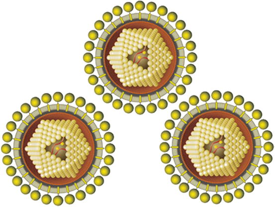
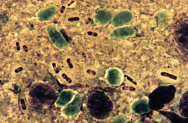
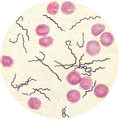
When studying by help of various techniques viruses (Fig. ).
Fig. 1.1
Diagrammatic representation of the tick-transmitted Spring-summer meningoencephalitis viruses measuring only 70 nm
Fig. 1.2
Light micrograph of the gram-negative plague bacteria ( Yersinia pestis ), which are transmitted by fleas and characterized by their bipolar staining. Their length is 1.50.5 m
Fig. 1.3
Light micrograph of a Giemsa stained blood smear showing stages of Borrelia recurrentis , the agent of tick-borne relapsing fever (measuring 8300.20.5 m)

