Melvin A. Shiffman and Alberto Di Giuseppe (eds.) Body Contouring Art, Science, and Clinical Practice 10.1007/978-3-642-02639-3_1 Springer-Verlag Berlin Heidelberg 2010
1. Mammary Anatomy
Michael R. Davis 1
(1)
Division of Plastic Surgery, University of Alabama, Birmingham School of Medicine, 510 20th Street South, 1164 Faculty Office Tower, Birmingham, AL 35294-3411, USA
Abstract
A thorough understanding of breast development and anatomy is a requirement for modern plastic surgeons. Advanced techniques of reduction mammaplasty, mastopexy, augmentation, and reconstruction demand a comprehensive knowledge of the current detailed descriptions of breast architecture. As a complicated physiologic and aesthetic structure, the form and function of the breast weighs heavily on a womans psyche. Significant improvements or complications can impact greatly on the self image for better or worse. Optimizing results and avoidance of complications takes root in the knowledge of breast anatomy. Only then can a plastic surgeon engage his full creativity in sculpting the breast form.
1.1 Introduction
A thorough understanding of breast development and anatomy is a requirement for modern plastic surgeons. Advanced techniques of reduction mammaplasty, mastopexy, augmentation, and reconstruction demand a comprehensive knowledge of the current detailed descriptions of breast architecture. As a complicated physiologic and aesthetic structure, the form and function of the breast weighs heavily on a womans psyche. Significant improvements or complications can impact greatly on the self image for better or worse. Optimizing results and avoidance of complications takes root in the knowledge of breast anatomy. Only then can a plastic surgeon engage his full creativity in sculpting the breast form.
1.2 Development (Fig. )
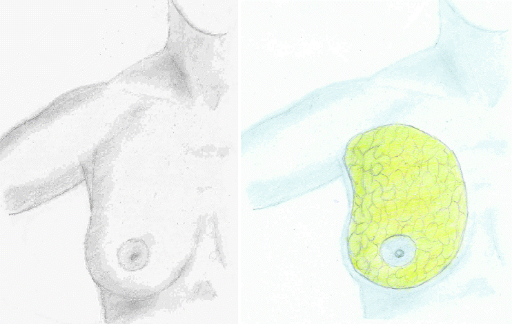
Fig. 1.1
The breast overlies the anterolateral chest wall containing primarily glandular tissue and fibrofatty stroma
As a cutaneous appendage, the breast takes its origin from the ectoderm. The breast bud begins differentiation during weeks 810 along the milk ridge. The normal human breast develops over the fourth intercostal space of the anterolateral chest wall. Supernumerary nipples and breasts can occur anywhere along the milk ridge from the axilla to the groin. Statistically, they are most common near the left inframmary crease.
Following a brief period of activity shortly after birth in response to maternal hormones, breast development becomes dormant until the onset of puberty. Pubertal onset is becoming ever earlier in modern society but currently occurs at approximately 9 years of age. Typically, by the age of 14, parenchymal growth has extended to its mature borders. These include the sternum medially, the anterior border of the latissimus dorsi laterally, the clavicle superiorly, and the inframammary crease inferiorly. These represent approximate anatomic landmarks and are not rigidly defined borders. Breast tissue can extend across the midline and beyond the inframammary crease. An extension of the breast tissue normally penetrates the axillary fascia into the axillary fat pad and is termed the Tail of Spence. Mature breast morphology projects off the chest wall in a conical fashion with its apex deep to the nippleareola complex.
Development of overall breast shape is multifactorial. Breast form is dependent on fat content and location, muscular and skeletal chest wall contour, and skin quality. These structures display complex attachments and interactions to result in the final form. Breast shape and size is unique to each individual and is determined largely by heredity.
1.3 Parenchyma (Fig. )
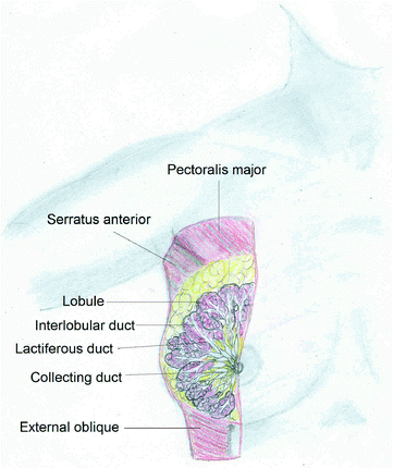
Fig. 1.2
Glandular breast tissue is lobular in structure with 2025 lobes each drained by a lactiferous duct. Milk then enters the collecting ducts followed by lactiferous sinuses prior to exiting the nipple
Embedded within the fibrofatty stroma lays the glandular portion of the breast. Glandular structure consists of millions of lobules clustered to comprise approximately 2025 lobes. Interlobular ducts come together to form approximately 20 main lactiferous ducts. Lactiferous sinuses collect milk, and specialized ducts within the nipple transmit milk to the surface. Glandular size remains relatively constant from individual to individual. The bulk of the breast consists of fat. Subcutaneous as well as interlobular fat content determine the texture, contour, and density.
The breast parenchyma is encompassed and supported by an intricate fascial system. The superficial fascial system is variable and sometimes indistinct from the overlying dermis anteriorly. Fat content of the subcutaneous tissue between the dermis and superficial fascia determines the clarity of these structures. Continuous with the superficial fascia is a deep component that separates the parenchyma from the pectoral fascia as well as the fascia overlying the adjacent muscles. Interposed between the superficial and deep components of the superficial fascial system are fascial extensions termed Coopers ligaments. Anchored to the muscular fascia, these ligaments act to suspend the parenchyma. Attenuation of these tissues is largely responsible for ptosis.
1.4 Musculature
At its foundation, the breast sits on a prominent musculature that also impacts form and physiology. The five primary muscle groups that lie deep into the breast are pectoralis major and minor, serratus anterior, upper external oblique, and upper rectus abdominis. Perforating these structures are the breasts primary arterial, venous, nerves, and lymphatic supply.
1.5 Skeletal Support
Breast symmetry and form is also dependent on normal skeletal support. The breast overlies the anterolateral thorax principally over ribs 26. Conditions which manifest chest wall abnormalities such as pectus excavatum and carinatum, Marfans syndrome, and Polands syndrome can present a challenge in optimizing breast aesthetics. It is also important to take note of the changes in the chest wall contour induced by plastic surgical intervention such as breast augmentation.
1.6 Arterial Supply (Fig. )
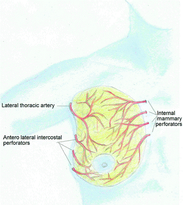
Fig. 1.3
Blood supply: The arterial supply to the breast is predominantly by perforators from the internal mammary artery followed by the lateral thoracic and anterolateral intercostals arteries
Breast tissue possesses a rich blood supply from multiple arterial sources. These sources collateralize within the breast to make a redundant system with significant clinical implications. Division of parenchyma is safe provided one of the several primary axes is preserved.
Entering the superomedial portion of the breast over intercostal spaces 26 are perforators from the internal mammary artery. These vessels supply the medial pectoralis muscle prior to entering the breast tissue and overlying skin. The dominant perforators emanate from the second and third intercostal spaces. These should be spared during reduction mammoplasty utilizing the superomedial pedicle. Of note, they are occasionally of adequate caliber for use as recipient vessels for free flap breast reconstruction.

