• Complain

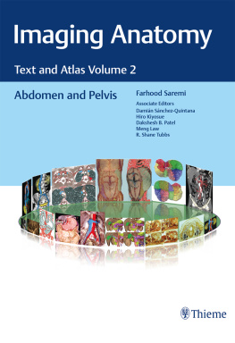
Farhood Saremi - Imaging Anatomy: Text and Atlas Volume 2: Abdomen and Pelvis

Here you can read online Farhood Saremi - Imaging Anatomy: Text and Atlas Volume 2: Abdomen and Pelvis full text of the book (entire story) in english for free. Download pdf and epub, get meaning, cover and reviews about this ebook. year: 2022, publisher: Thieme Medical Publishers, genre: Romance novel. Description of the work, (preface) as well as reviews are available. Best literature library LitArk.com created for fans of good reading and offers a wide selection of genres:

Romance novel Science fiction Adventure Detective Science History Home and family Prose Art Politics Computer Non-fiction Religion Business Children Humor

Choose a favorite category and find really read worthwhile books. Enjoy immersion in the world of imagination, feel the emotions of the characters or learn something new for yourself, make an fascinating discovery.

Farhood Saremi Imaging Anatomy: Text and Atlas Volume 2: Abdomen and Pelvis

Imaging Anatomy: Text and Atlas Volume 2: Abdomen and Pelvis: summary, description and annotation

We offer to read an annotation, description, summary or preface (depends on what the author of the book "Imaging Anatomy: Text and Atlas Volume 2: Abdomen and Pelvis" wrote himself). If you haven't found the necessary information about the book — write in the comments, we will try to find it.

Unique anatomic atlas provides an indispensable virtual desk dissection experience

Normal imaging anatomy and variants, including diagnostic and surgical anatomy, are the cornerstones of radiologic knowledge. Imaging Anatomy: Text and Atlas Volume 2, Abdomen and Pelvis is the second in a series of four richly illustrated radiologic references edited by distinguished radiologist Farhood Saremi. The atlas is coedited by esteemed colleagues Damin Snchez-Quintana, Hiro Kiyosue, Dakshesh B. Patel, Meng Law, and R. Shane Tubbs with contributions from an impressive cadre of international authors. Succinctly written text and superb images provide readers with a virtual, user-friendly dissection experience.

This exquisitely crafted atlas combines fundamental core anatomy principles with modern imaging and postprocessing methods to increase understanding of intricate anatomical features. Twenty-two concise chapters cover the abdominal wall, alimentary tract, liver, biliary system, pancreas, spleen, peritoneum, genitourinary system, pelvic floor, neurovasculature, and surface anatomy. Relevant anatomical components of the abdomen and pelvis are discussed, including musculature, arteries, veins, lymphatics, ducts, and innervation.

Key Highlights

  • High-quality cross-sectional multiplanar and volumetric color-coded CT, MRI, and angiography imaging techniques provide detailed insights on specific anatomy
  • Cross-sectional and topographic cadaveric views by internationally known anatomists coupled with more than 1,600 illustrations clearly elucidate difficult anatomical concepts
  • Consistently formatted chapters include an introduction, embryology, review of anatomy, discussion of anatomical variants, postsurgical anatomy, and congenital and acquired pathologies

This unique resource provides an excellent desk reference for differentiating normal versus pathologic anatomy. It is essential reading for medical students, radiology residents and veteran radiologists, internists, and general surgeons, as well as vascular and transplant surgeons.

This print book includes complimentary access to a digital copy on https://medone.thieme.com.

Publishers Note: Products purchased from Third Party sellers are not guaranteed by the publisher for quality, authenticity, or access to any online entitlements included with the product.

Farhood Saremi: author's other books


Who wrote Imaging Anatomy: Text and Atlas Volume 2: Abdomen and Pelvis? Find out the surname, the name of the author of the book and a list of all author's works by series.

Imaging Anatomy: Text and Atlas Volume 2: Abdomen and Pelvis — read online for free the complete book (whole text) full work

Below is the text of the book, divided by pages. System saving the place of the last page read, allows you to conveniently read the book "Imaging Anatomy: Text and Atlas Volume 2: Abdomen and Pelvis" online for free, without having to search again every time where you left off. Put a bookmark, and you can go to the page where you finished reading at any time.

Light

Font size:

Reset

Interval:

Bookmark:

Make
Imaging Anatomy Text and Atlas Volume 2 Abdomen and Pelvis Farhood Saremi MD - photo 1

Imaging Anatomy

Text and Atlas Volume 2
Abdomen and Pelvis

Farhood Saremi, MD
Professor of Radiology and Medicine
Department of Radiology
University of Southern California
Keck Medicine of USC
Los Angeles, California, USA

Associate Editors:

Damin Snchez-Quintana, MD, PhD
Professor of Human Anatomy
Faculty of Medicine
Department of Anatomy and Cell Biology
University of Extremadura
Badajoz, Spain

Hiro Kiyosue, MD, PhD
Professor of Diagnostic Imaging and Analysis
Faculty of Life Sciences
Kumamoto University
Kumamoto, Japan

Dakshesh B. Patel, MD
Associate Professor of Clinical Radiology
Department of Radiology
University of Southern California
Keck Medicine of USC
Los Angeles, California, USA

Meng Law, MD

Professor of Radiology, Neurology, and Neurological Surgery

University of Southern California;

Biomedical Engineering

Viterbi School of Engineering;

Director of Neuroradiology and the Neuroradiology Fellowship Program

Keck Medicine of USC

Los Angeles, California, USA

R. Shane Tubbs, MD

Professor, Chief Scientific Officer, and Vice President

Seattle Science Foundation

Seattle, Washington, USA

1612 illustrations

Thieme
New York Stuttgart Delhi Rio de Janeiro

Library of Congress Cataloging-in-Publication Data is available from the publisher

2023. Thieme. All rights reserved.

Thieme Medical Publishers, Inc.

333 Seventh Avenue, 18th Floor,

New York, NY 10001, USA

.thieme.com

+1 800 782 3488,

Cover design: Thieme

Cover images source: Thieme

Typesetting by Thomson Digital, India

Printed in Germany by Beltz Grasche Betriebe 5 4 3 2 1

ISBN: 978-1-62623-982-1

Also available as an e-book:

eISBN (PDF): 978-1-62623-983-8

eISBN (epub): 978-1-63853-611-6

Important note: Medicine is an ever-changing science undergoing continual development. Research and clinical experience are continually expanding our knowledge, in particular our knowledge of proper treatment and drug therapy. Insofar as this book mentions any dosage or application, readers may rest assured that the authors, editors, and publishers have made every effort to ensure that such references are in accordance with the state of knowledge at the time of production of the book.

Nevertheless, this does not involve, imply, or express any guarantee or responsibility on the part of the publishers in respect to any dosage instructions and forms of applications stated in the book. Every user is requested to examine carefully the manufacturers leaflets accompanying each drug and to check, if necessary in consultation with a physician or specialist, whether the dosage schedules mentioned therein or the contraindications stated by the manufacturers differ from the statements made in the present book. Such examination is particularly important with drugs that are either rarely used or havebeennewly released on the market. Every dosage schedule or every form of application used is entirely at the users ownrisk and responsibility. The authors and publishers request every user to report to the publishers any discrepancies or inaccuracies noticed. If errors in this work are found after publication, errata will be posted at www.thieme.com on the product description page.

Some of the product names, patents, and registered designs referred to in this book are in fact registered trademarks or proprietary names even though specific reference to this fact is not always made in the text. Therefore, the appearance of a name without designation as proprietary is not to be construed as a representation by the publisher that it is in the public domain.

Thieme addresses people of all gender identities equally. We encourage our authors to use gender-neutral or gender-equal expressions wherever the context allows.

This book, including all parts thereof, is legally protected by copyright. Any use, exploitation, or commercialization outside the narrow limits set by copyright legislation without the publishers consent is illegal and liable to prosecution. This applies in particular to photostat reproduction, copying, mimeographing or duplication of any kind, translating, preparation of microfilms, and electronic data processing and storage.

Contents

Farhood Saremi

Preface

In the preface of his anatomy text collection de Humani Corporis Fabrica published in 1543, Andreas Vesalius wrote anatomy should rightly be regarded as the strong foundation of the whole art of medicine , acknowledging the fundamental role the study of anatomy has in the practice of medicine. Furthermore, one of the major lasting successes of his influential anatomy series was the ability to reproduce many superb illustrations of human anatomy dissection which was made possible by technical developments during his time.

Likewise, recent advancement in imaging technology and the development of newer postprocessing software have increased our ability to image and show anatomic detail of the human organs. At the same time, over the past decade, the literature is filled with many precious articles on anatomy and related topics. Unfortunately, there is an increasing gap between our fundamental core anatomy reference texts and our understanding and depiction of human anatomy using modern imaging and postprocessing methods. The idea for this project arose from the necessity for a text to fill this gap and describe anatomy in the context of current advances in imaging technology and science.

Computed tomography (CT) and magnetic resonance (MR) have been the traditional methods in performing noninvasive studies, and both have immensely contributed to our ability to deliver accurate diagnostic information. Current generation MR and CT as well as dedicated peripheral and intraluminal ultrasound provide the spatial and contrast resolutions required to demonstrate anatomic details with unprecedented accuracy.

When it comes to assessment of details, there is no imaging modality that can compete with the speed, accuracy, and spatial resolution of new CT scanners. Volume rendering and mutliplanar reformations facilitate understanding of the complex structures such as heart, vessels, and bones. With current scanners, the entire anatomic span of the major organs can be covered in a few seconds with spatial resolution of less than 0.5 mm. In this series, this technology along with the state-of-the-art postprocessing methods have been used to create volumetric color-coded images.

On the other hand, MRI provides high levels of contrast resolution that would be difficult to obtain with CT. High-field MRI provides superb resolution of brain anatomy. Taking advantage of a 7 Tesla MR scanner at our facility, high-quality images were obtained to complete the chapters of the neuroanatomy volume (volume 4). Using MR tractography, it is possible to depict the anatomic course of the white tracts in detail, and with new postprocessing software it is further possible to map areas of the brain cortex and subfield regions of small structures such as hippocampus. These technologies have ushered in a new era for investigation of brain anatomy and are being used for the planning of sophisticated brain surgeries and for localization of small regions of interest.

This imaging anatomy series is a reference atlas and text with a practical discussion of specific topics along with examples of anatomical variants. Divided into four major volumes, each volume takes the instructive format of a classic text but is infused with a large number of images and illustrations to teach readers anatomy via state-of-the-art cross-sectional and volumetric imaging. The volumes are divided into lung, mediastinum, and heart; abdomen and pelvis; musculoskeletal; and head, neck, and spine imaging. Peripheral vasculature and nerves have been included in the musculoskeletal volume.

Next page
Light

Font size:

Reset

Interval:

Bookmark:

Make

Similar books «Imaging Anatomy: Text and Atlas Volume 2: Abdomen and Pelvis»

Look at similar books to Imaging Anatomy: Text and Atlas Volume 2: Abdomen and Pelvis. We have selected literature similar in name and meaning in the hope of providing readers with more options to find new, interesting, not yet read works.


Reviews about «Imaging Anatomy: Text and Atlas Volume 2: Abdomen and Pelvis»

Discussion, reviews of the book Imaging Anatomy: Text and Atlas Volume 2: Abdomen and Pelvis and just readers' own opinions. Leave your comments, write what you think about the work, its meaning or the main characters. Specify what exactly you liked and what you didn't like, and why you think so.