Z. M. Seagal - Atlas of Topographical and Pathotopographical Anatomy of the Head and Neck
Here you can read online Z. M. Seagal - Atlas of Topographical and Pathotopographical Anatomy of the Head and Neck full text of the book (entire story) in english for free. Download pdf and epub, get meaning, cover and reviews about this ebook. year: 2018, publisher: Wiley-Scrivener, genre: Science. Description of the work, (preface) as well as reviews are available. Best literature library LitArk.com created for fans of good reading and offers a wide selection of genres:
Romance novel
Science fiction
Adventure
Detective
Science
History
Home and family
Prose
Art
Politics
Computer
Non-fiction
Religion
Business
Children
Humor
Choose a favorite category and find really read worthwhile books. Enjoy immersion in the world of imagination, feel the emotions of the characters or learn something new for yourself, make an fascinating discovery.
- Book:Atlas of Topographical and Pathotopographical Anatomy of the Head and Neck
- Author:
- Publisher:Wiley-Scrivener
- Genre:
- Year:2018
- Rating:4 / 5
- Favourites:Add to favourites
- Your mark:
Atlas of Topographical and Pathotopographical Anatomy of the Head and Neck: summary, description and annotation
We offer to read an annotation, description, summary or preface (depends on what the author of the book "Atlas of Topographical and Pathotopographical Anatomy of the Head and Neck" wrote himself). If you haven't found the necessary information about the book — write in the comments, we will try to find it.
Z. M. Seagal: author's other books
Who wrote Atlas of Topographical and Pathotopographical Anatomy of the Head and Neck? Find out the surname, the name of the author of the book and a list of all author's works by series.

Atlas of Topographical and Pathotopographical Anatomy of the Head and Neck — read online for free the complete book (whole text) full work
Below is the text of the book, divided by pages. System saving the place of the last page read, allows you to conveniently read the book "Atlas of Topographical and Pathotopographical Anatomy of the Head and Neck" online for free, without having to search again every time where you left off. Put a bookmark, and you can go to the page where you finished reading at any time.
Font size:
Interval:
Bookmark:
Z. M. Seagal is an Honoured Scientist of the Russian Federation and the Udmurt Republic, Honorary Academician of the Izhevsk State Medical Academy, Head of the Department of Operative Surgery and Topographic Anatomy, Doctor of Medical Sciences, and Professor.
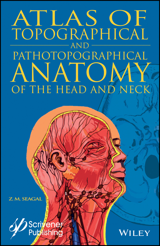
Topographical and Pathotopographical Medical Atlas of the Head and Neck.
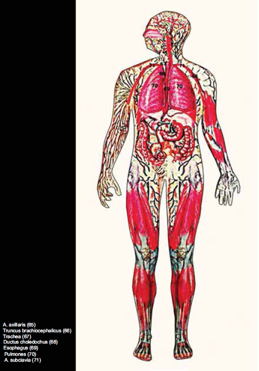
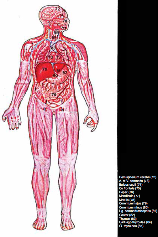
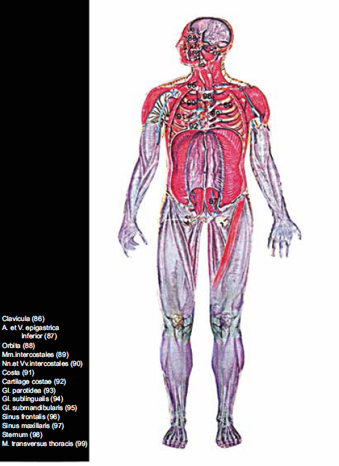
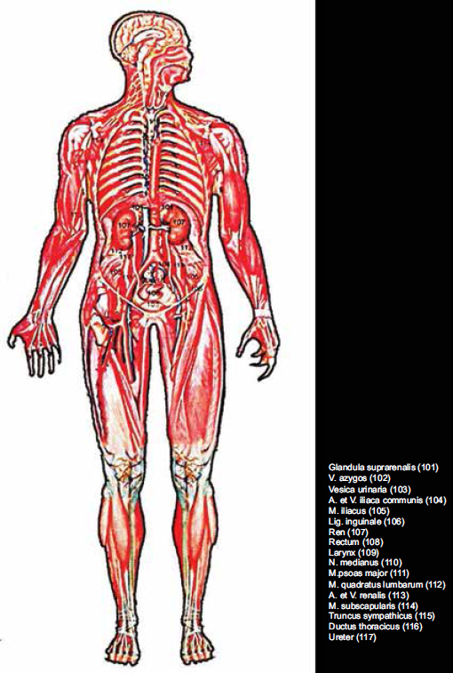
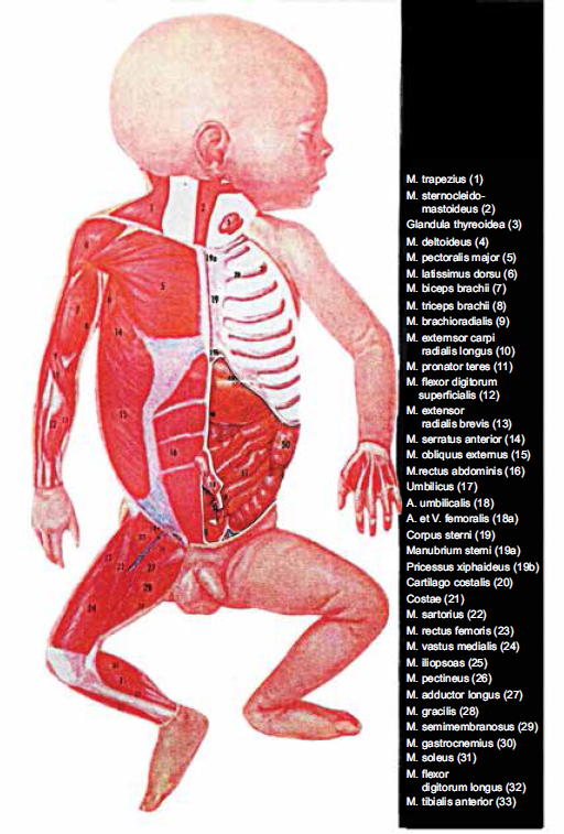
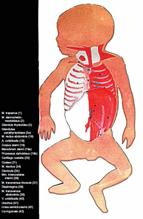
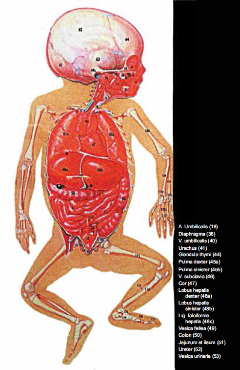
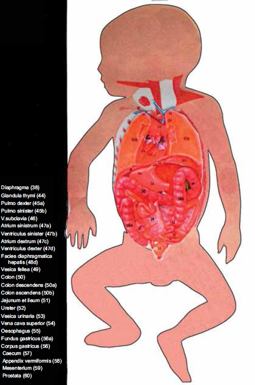
The atlas presents topographical and pathotopographic human anatomy for both the adult and the child. The section Head includes cerebral and facial parts according to the areas. Layer topographic anatomy, variant, computer and MRI-topographic anatomy are presented in the section. The surgical anatomy of congenital malformations of the head includes cerebral hernia and hydrocephalus, and of the face - macrostomy, coloboma, clefts of hard and soft palate. In the section Neck there are given individual and age differences, fasciae and cell spaces, triangles and vascular-neural bundles, collateral blood supply of the brain in injuries or occlusion of the main neck arteries, topography of the neck organs. All the pictures are colorful and original. The atlas is written in accordance with the educational program of medical universities of the Russian Federation. The original graphs of logical structures are presented according to the sections of topography and congenital malformations. This allows an effective study of the subject.
This atlas is intended for the students of General Medicine, Pediatrics and Dentistry faculties, as well as for interns, residents, postgraduate students and surgeons. The monograph is intended to be used by physicians, junior physicians, medical residents, lecturers in medicine, and medical students.
Ultrasonic Topographical and Pathotopographical Anatomy: A Color Atlas, by Z. M. Seagal and O. V. Surnina, ISBN 9781119223573. Using ultrasonic technology to create full-color detailed pictures, this atlas details the topographical and pathotopographical anatomy of the human body, as a useful reference for medical professionals and students alike. NOW AVAILABLE!
Compendium of Biophysics, by Andrey B. Rubin, ISBN 9781119160250. The most thorough coverage of biophysics available, in a handy, easy-to-read volume, perfect as a reference for experienced engineers or as a textbook for the novice. NOW AVAILABLE!
Fundamentals of Biophysics, by Andrey B. Rubin, ISBN 9781118842454. The most up-to-date and thorough textbook on the fundamentals of biophysics, for the student, professor, or engineer. NOW AVAILABLE!
Ethics in the University, by James G. Speight, ISBN 9781118872130. Examining the potential for unethical behavior by all academic staff, both professionals and nonprofessionals, this groundbreaking new study uses documented examples to show where the matter could have been halted before it became an ethics issue and how to navigate the maze of todays sometimes confusing ethical academic arena. NOW AVAILABLE!
Reactive Oxygen Species: Signaling Between Hierarchical Levels in Plants, by Franz-Josef Schmitt and Suleyman I. Allakhverdiev, ISBN 9781119184881. Not available anywhere else, this groundbreaking volume presents a novel and unique approach to understand the complex chemical network behind the internal network structure of plants, a valuable tool for students of physics, biological physics, chemistry and biology, but also for sociologists and economists as well as for scientists in any field. NOW AVAILABLE!
The Head
Limits, outer orienteers. The head is separated from the neck with the line which begins on chin elevation protuberantia mentalis, after that it laterally continues across the lower mandible edge, continues by the lower semicircle of outer aural meatus, goes on to the upper nuchal line, linea nuchae superior, and ends on both sides on outer elevation of occipital bone with protuberantia occipitalis externa. In general, the head is divided into cerebral cranium cranium cerebrale and facial cranium cranium faciale.
Limits: The cerebral cranium is separated from facial cranium with the anatomical formations described below. It is limited by glabella by the middle line. Afterwards this line goes along the brow arch, arcus superciliaris, then - by the rear edge of the processus zygomaticus ossis frontalis and processus frontosphenoidalis ossis zygomatici, then the line goes on by the zygomatic arch, then it continues vertically to the rear edge of the ascending branch of the mandible, and afterwards it continues on the bottom margin where it ends after connecting with a similar line coming from the opposite side forming protuberantia mentalis.
Area division: The cerebral cranium can be divided into the cranium base, basis cranii, and the cranium fornix (or dome) which is also called calvaria. The cranium base is divided into inner one (basis cranii interna) and external one (basis cranii externa). Calvaria is divided into frontal, parietal, occipital, temporal and mastoid regions (regg. Frontalis, parietalis, occipitalis, temporalis et mastoidea).
Regio frontalis (or the frontal area) is located within the frontal region. Its limiting line begins in the lower part of glabella, spreads to the sides across eyebrow arches, crosses the zygomatic process of the frontal bone, continues upwards across temporal line, and then ascends by the projection line of the coronal suture.
Regio parietalis (parietal region). Limits: anterior limit is the coronal suture, lambdoid suture is the rear limit, temporal line limits it from the sides.
Regio occipitalis (occipital region) is located within the squama of the occipital bone. Limits: lambdoid suture limits it from the top and from the sides and the line drawn horizontally from one apex of mastoid bone to another limits it from below. Its called linea bimastoidea.
Font size:
Interval:
Bookmark:
Similar books «Atlas of Topographical and Pathotopographical Anatomy of the Head and Neck»
Look at similar books to Atlas of Topographical and Pathotopographical Anatomy of the Head and Neck. We have selected literature similar in name and meaning in the hope of providing readers with more options to find new, interesting, not yet read works.
Discussion, reviews of the book Atlas of Topographical and Pathotopographical Anatomy of the Head and Neck and just readers' own opinions. Leave your comments, write what you think about the work, its meaning or the main characters. Specify what exactly you liked and what you didn't like, and why you think so.