Thomas Krieg , David R. Bickers and Yoshiki Miyachi (eds.) Therapy of Skin Diseases A Worldwide Perspective on Therapeutic Approaches and Their Molecular Basis 10.1007/978-3-540-78814-0_1 Springer-Verlag Berlin Heidelberg 2010
11. Biology of the Skin
The skin is the largest organ of the body and crucial for terrestrial life by providing a sturdy barrier toward the outside world. This barrier protects the organism from dehydration and prevents microbes and damaging agents from entering. The skin is challenged on a daily basis by a range of external insults, such as changes in temperature, UV light, and bacteria to thermal and mechanical injuries. Since in most cases, the skin is able to handle challenges without the occurrence of overt disease, this organ, by nature, must be an extremely versatile and dynamic tissue.
During evolution, the skin has gained a number of structural and functional features that allow it to react in an adequate manner to those external signals and injuries. Most importantly, the skin has developed ultra-structurally defined subcompartments that topically restrict external and internal damage.
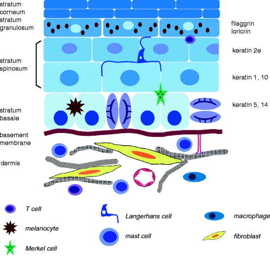
The skin is composed of an epithelial and a mesen-chymal compartment, the epidermis and the dermis, which are connected by a highly specialized extracellular matrix structure, and the basement membrane zone (Fig. ). The dermis is resting on the subcutis, a fat layer that connects it to the fascia and the underlying muscles. These two compartments communicate extensively in various ways and at different levels, and this is crucial to establish, maintain, and restore homeo-stasis. During skin morphogenesis, this reciprocal interaction also determines the formation of the epidermal appendages, such as hair follicles, sweat glands, sebaceous glands, and nails, all structures that are required for normal skin function. Strong regional differences exist in the thickness and differentiation status of dermis and/or epidermis and the distribution of skin appendages. These variations are ontogenetically determined and form the basis for differential skin function required in various anatomical areas.
Fig. 1.1.1
Schematic representation of human skin. This simplified model depicts the major cell types present in the skin under homeostatic conditions. Their presence and number vary depending on the anatomical location of the skin. Note that hair follicles and other appendages are not represented
This chapter provides a general overview of, and introduction to, the cellular composition of the skin, the most important functions of the different cell types, and their particular contribution to the multifunctional skin barrier. Although it emphasizes several aspects more than others, this chapter does not aim at going extensively into details, and for the most part, uses citations of excellent and comprehensive reviews to refer to the reader who is interested to learn more on particular subjects. In addition, many chapters of this book will discuss the different aspects discussed here in light of skin disease.
11.1 Cellular Composition of the Epidermis
The epidermis and its appendages, hair follicles, sebaceous, and sweat glands form the physical barrier of the organism of the outside world. As a barrier, it serves several important functions, both physical and immunological, which are reflected in the cell types and differentiation status that make up the epidermis.
Keratinocytes are the most predominant cell type in the epidermis and form the cornerstone of its overall structure and function. Epidermal keratinocytes balance lifelong self-renewal with a spatiotemporally strictly regulated terminal differentiation program, which ultimately leads to the formation of a dead, cornified, and water impermeable cell layer []
Different populations of stem and progenitor cells located in the basal layer of interfollicular epidermis (IFE) and in specific areas of hair follicles guarantee constant self-renewal under steady state conditions and sufficient plasticity for the fast replacement of lost tissue in case of injury. Morphogenetic signal pathways, such as Wnts, BMPs/TGF- , Notch and Hedgehogs, control the determination, renewal and maintenance of these stem cells []. The flexible balance between self-renewal and terminal differentiation is determined by the variable conditions of the extracellular environment.
Over the last decade, it has become clear that kera-tinocytes also actively contribute to the immunological barrier of the skin []. These cells produce antimicrobial peptides, such as defensins, and express Toll-like receptors on their cell surface, important for the control of innate immunity. In addition, upon disturbance of skin homeostasis, these cells secrete a wide range of cytokines and other growth factors that influence the innate and adaptive immune response.
Although over 90% of the epidermis consists of keratinocytes, other cells types, such as Langerhans cells, T-cells, melanocytes and Merkel cells, are present in the different layers (Fig. ). These cells serve crucial specialized functions that contribute to epidermal homeostasis and to its restoration upon challenges of the epidermal barrier.
Merkel cells are postmitotic, neuroendocrine cells that produce a large number of cytokines and neuro-peptides, form close connections with sensory nerve endings, and are mainly located in the basal layer of the epidermis. Ultrastructurally, these cells are characterized by dense core granules. Although these cells are the least well characterized cells in the epidermis, they are thought to have mechanosensory functions and contribute to the regulation of inflammatory responses []
Langerhans cells are immature dendritic cells that form close contacts with keratinocytes and monitor microbial infection. Upon activation, these cells mature and migrate to draining lymph nodes where they present antigens to T-lymphocytes []
The epidermis also contains a resident population of unique T-cells, which are in close contact with Langerhans cells and keratinocytes. These cells regulate skin inflammatory responses and play important roles in graft vs. host reactions in the skin []. The production and transfer of melanosomes is a complex and incompletely understood process, but plays a crucial role in the defense against the daily assault of UV light.
11.2 Cellular and Structural Composition of the Dermis
In contrast to the epidermis, the dermis is rich in extracellular matrix (ECM), and contains relatively fewer cells (Fig. ). The upper or papillary dermis is characterized by loose connective tissue and a horizontal plexus of blood vessels, which are connected to a deep plexus located in the subcutis. The lower or reticular dermis makes up the major part of the dermis and mainly contains thick collagen bundles.


