Springer International Publishing Switzerland 2016
Bertrand Richert , Nilton Di Chiacchio , Marie Caucanas and Nilton Gioia Di Chiacchio Management of Ingrowing Nails 10.1007/978-3-319-30555-4_1
1. Basics
Abstract
Performing successful nail surgery requires a comprehensive knowledge of nail anatomy and physiology. Understanding both the vascular and neural pathways supplying the nail complex, the functions and relationship of each component of the nail unit is also essential. The preoperative consultation is mandatory as it allows a detailed evaluation of the patient as well as a full information about the procedure and post operative evolution. The nail surgeon should use adequate instruments for nail surgery and be aware of the techniques and tips allowing an efficient anesthesia with minimal patient discomfort. Post operative procedure includes the proper use of painkillers and wound care. As for any kind of surgery, the operator needs to prevent and handle any complication that may occur during follow-up.
Surgical Anatomy
Introduction
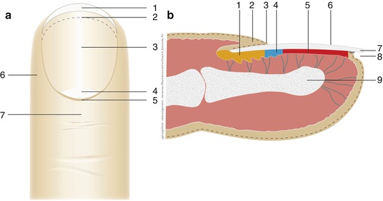
Before embarking to any nail surgery, a good knowledge of the anatomy of the nail unit is mandatory (Fig. ). It will help to understand how to perform an adequate local anesthesia as well as reasonable procedures at that site and how to deal with post operative bleeding.
Fig. 1.1
( a ) Anatomy of the nail apparatus. Upper view. free edge, hyponychium, nail bed. lunula, cuticle, lateral nail fold, proximal nail fold. ( b ) Anatomy of the nail apparatus. Lateral view. proximal nail matrix, proximal nail fold, cuticle, distal nail matrix, nail bed, nail plate, free edge, distal groove, distal phalanx
The nail apparatus is an integral part of the tip of the digit. All parts are intimately related to each other forming a functional, sensory and cosmetic unit. It is made up of the distal bony phalanx with the joint and synovial membrane, a fibrous network consisting of ligaments, tendons, and connective tissue strings, blood vessels and glomus bodies, nerves and receptors making it an extremely efficient sensory organ and the nail unit [].
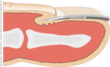
Nail Matrix
The nail matrix constitutes the sole germinative structure responsible for the production of the nail plate. It is located on the proximal dorsal aspect of the distal phalanx and just distal to the interphalangeal joint, mostly covered by the proximal nail fold (PNF). The matrix rests on the base of the distal bony phalanx and forms a crescent with posterior inferior concavity (Fig. ].
Fig. 1.2
( a ) Anatomic position of the nail matrix. Upper and transverse view. ( b ) Anatomic position of the nail matrix on the great toenails. Lateral view
Fig. 1.3
Formation of the nail plate by the matrix: the superficial upper third comes from the proximal matrix, the lower 2/3 from the distal matrix
Fig. 1.4
( a ) Longitudinal rete ridges running on the whole length of the nail bed. ( b ) Undersurface of the nail plate showing the complementary set of ridges (after friction with ink)
Key Points
The lateral horns of the matrix may extend up and even beyond the midline of the lateral aspect of the great toenail .
Nail Bed
The nail bed (also called sterile matrix by some surgeons as it does not produce any nail substance) is the distal continuation of the nail matrix. It corresponds to the pinkish area that spreads from the distal lunula border till the hyponychium. It very firmly adheres to the nail plate. On the nail bed runs a unique structure that is responsible for its very firm adherence to the plate: longitudinal parallel rete ridges, often very clearly observed after nail avulsion (Fig. ].
Nail Folds
The nail unit is surrounded on three sides by the proximal and lateral nail folds in an inverse U shape, constituting the paronychium. The PNF protects and covers the nail matrix and the newly formed nail plate. The proximal nail groove is a pocket formed by the PNF, and its proximal part is called cul-de-sac. Its dorsal surface is made from a skin with a thin dermis and almost no appendages. Some sweat glands are seen in its most proximal portion. The acute angle formed by the free margin of the PNF is essential for the formation of the cuticle. The cuticle is divided into two parts: the true cuticle, attached to the underlying newly formed nail and produced by the deep portion of the ventral surface, and the false cuticle, formed by the horny layers of the dorsal roof and the distal third of the ventral surface of the PNF [].
The lateral nail folds are connective tissue rolls that progressively flatten from the PNF to the distal tip of the digit. They are often very pronounced in the lesser toes and sometimes in the great toenails rendering some patients prone to develop ingrowing nails. The lateral grooves are longitudinal parallel indentations framing the lateral nail margins and providing an abutment for the nail, for which they have a specialized connective tissue arrangement [].
Hyponychium
The hyponychium is the transition of the nail bed to the digital pulp. It has, like the cuticle, a sealing function. At its most distal part, the nail plate separates from the nail bed. The hyponychial attachment is seen through the nail as the onychodermal band [].
Nail Plate
The nail plate is synthesized by the matrix. It is composed of compacted keratinized epithelial cells, continuously and exclusively formed by the nail matrix. It is a unique combination of strength and flexibility. The thin dorsal layer of the nail plate (upper third) produced by the proximal matrix, has a smooth shiny surface and is made up of many strongly flattened onychocytes with considerable intercellular adhesion structures. The intermediate layer originates from the distal matrix and is much thicker, but its cells are less flattened than those of the superficial layer. The ventral layer is mainly nail bed (onycholemmal) keratin and has longitudinal ridges that correspond to complementary ridges on the upper aspect of the nail bed. These nail ridges may be best examined using polarized light.
Protection and enhancement of the sensory functions of the fingertip are the most important functions of several of the nail plate. While the toenails are formed over a period of 1218 months, the fingernails grow continuously on an average of 0.1 mm per day. As well as the toenails, the nails of the shorter fingers grow comparatively slower.