1. Introduction: Anatomy of the Lips and Eye
1.1 Anatomy of the Eyelids
The eyelids are highly specialized structures with peculiar anatomic components. The ocular globes are allocated in two symmetrically bony cavities called orbits, consisting of seven bones that develop the orbital walls. The roof is composed mostly of the orbital plate of the frontal bone and posteriorly of a minor part of the sphenoid bone. The lateral wall comprises the orbital surface of the zygomatic bone and the sphenoid bone. The floor is composed of the orbital plate of the maxilla anterolaterally of the zygomatic bone and posteriorly of the palatine bone. The medial wall consists of the ethmoid, frontal, lacrimal, and sphenoid bone. The eyelid skin, which is less than 1 mm thick, is a thin epidermis constructed from a stratified epithelium of 67 cell layers. The dermis contains elastic fibers, blood vessels, lymphatics, and nerves. The underlying fat is scant or not present in the subcutaneous tissue, where the hair follicles and pilosebaceous glands are located. The apocrine glands of Moll are located near the lid margin, and the sebaceous glands of Zeiss are associated with the follicles of the eyelashes. The eyelids function to protect the eye globe from local and external injuries. Furthermore, they regulate the light that reaches the eye and uniformly distribute the tear film, mucus, and oil during blinking, of great importance for the health of the cornea. The eyelids are divided into upper and lower eyelids, which are similar but with different characteristics mainly in the lid retractor arrangement. The space between the open lids is known as the palpebral fissure, which measures 712 mm, while the normal excursion of the lids is 1417 mm. In the normal adult fissure, the highest point of the upper lid is just nasal to the center of the pupil, while the lowest point of the lower lid is just temporal to the center of the pupil. In youths, the upper lid margin rests at the upper limbus, whereas in adults it rests 1.5 mm below the limbus. The lower eyelid margin rests at the level of the lower limbus. The lateral canthal angle is 2 mm higher than the medial canthal angle in Europeans, but is 3 mm higher in Asians. The distance from the medial canthus to the midline of the nose is approximately 15 mm. The lateral canthus lies directly over the sclera, and the medial canthus is separated from the eye by the lacrimal lake and caruncle, a yellowish tissue containing sebaceous and sweat glands. The lid margins are 2 mm wide and form the junction between the skin and the conjunctiva, the mucous membrane of the lids. They meet at the gray line, near the posterior edge of the lid margin, the junction of the anterior and posterior lamellae of the lids. The eyelashes are located anteriorly and the openings of the meibomian glands posteriorly. There are approximately 100150 eyelashes on the upper lid and about 5075 on the lower. The follicular structure of eyelashes includes the sebaceous (Zeiss) and sweat (Moll) glands, while the tarsal glands (Meibomian) open posteriorly to the lid margin. The tears that appear at the tips of the small papillae are drained from the surface of the eyes through the openings by a pump mechanism. The lacrimal secretory system controls the amount of tears and is divided into the basic and reflex secretors. The basic secretor is composed of three sets of glands. (1) Conjunctival, tarsal, and limbal mucin-secreting goblet cells; the overlying aqueous layer is spread more uniformly because of this inner layer (precorneal tear film). (2) The accessory lacrimal exocrine glands of Krause and Wolfring, located in the subconjunctival tissue. (3) The oil-producing Meibomian glands and the palpebral glands of Zeiss and Moll. The reflex secretor is divided into two parts by the lateral horn of the levator palpebrae superioris.
The first fold of the upper eyelid is represented by the superior palpebral sulcus, 910 mm (individual and racial variations) above the lid margin, and represents the junction of the levator palpebrae superioris with the orbital septum and the fibrous insertion of the levator aponeurosis into the skin.
There is a thin fascial layer between the skin and the orbicularis oculi muscle, with no fat tissue. The eyelid normally is located at the superior border of the tarsus, and the skin below the lid is attached to the underlying tarsus with the levator aponeurosis, which has projections anteriorly through the pretarsal orbicularis to the skin and posteriorly to the inferior portion of the anterior tarsus. The skin of the upper eyelid is more freely movable because of the lack of superior aponeurotic attachments and underlying orbital septum.
The second layer of the eyelid is the orbicularis oculi muscle, which is divided into orbital and palpebral parts that function independently. The orbital part is a voluntary muscle while the palpebral part is both voluntary and involuntary. The orbital portion extends in a wide, circular fashion around the orbit, interdigitating with other muscles of facial expression. It has a curved origin from the medial orbital margin, being attached to the superomedial orbital margin, maxillary process of the frontal bone, medial palpebral ligament, frontal process of the maxilla, and inferomedial orbital margin. The palpebral portion is further subdivided into pretarsal and preseptal portions. The preseptal orbicularis muscle covers the orbital septum and originates medially from a superficial and deep head associated with the medial palpebral ligament. The fibers from the upper and lower eyelid join laterally to form the lateral palpebral raphe, which is attached to the overlying skin. The pretarsal portion lies anterior to the tarsus, with a superficial and deep head of origin intimately associated with the medial palpebral ligament. Fibers run horizontally and laterally to extend deep to the lateral palpebral raphe, to insert in the lateral orbital tubercle through the intermediary of the lateral canthal tendon. The peripheral fibers sweep across the eyelid over the orbital margin in a series of concentric loops, the more central ones forming almost complete rings, interdigitating with other muscles of facial expression. In the upper lid the orbital part extends as far as the forehead, covers the corrugator supercilii muscle, and continues laterally over the anterior temporal fascia.
The third layer of the lids in the upper portion is the orbital septum, a fascial membrane that separates the eyelid structures from the deeper orbital structures, and attaches to the orbital margin a thickening called the arcus marginalis, the point of confluence for the facial bone periosteum and the periorbita. With age, the septum weakens and bulging of the orbital fat pad becomes visible. Its removal is important in blepharoplastic surgery.
The fourth layer of the upper lid is the postseptal fat pad, contained within the orbit by the orbital septum.
In the lower lid, the orbital part lies on the origins of the elevator muscles of the upper lip and nasal ala, and continues to cover partially the masseter muscle (Figs. ).
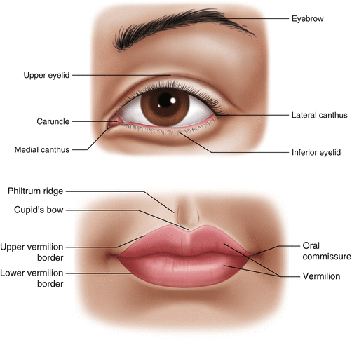
Fig. 1.1
Eyelids: () Eyebrow, () Upper eyelid, () Medical cantus, () Caruncle, () Lateral cantus, () Inferior eyelid. Lips: () Philtrum ridge, () Cupids bow, () Upper vermilion border, () Vermilion, () Oral commessure, () Lower vermilion border