Introduction
Sound working knowledge of the bones, muscles, vasculature, innervation, fat depots, and retaining ligaments of the face is the cornerstone to precise, reproducible, and aesthetic surgical results. In the midface, intimate familiarity with these structures is even more crucial, given the continuous fibromuscular sheath that encases all of the muscles of facial expression in this area. Therefore, contraction of one muscle engages the overlying skin of the entire midface, leading to facial expression. The midface is crucial for facial expression, eyelid closure, eyelid position, and speech articulation.
Aesthetically, the malar folds, nasojugular folds, and jowls all stem from senescence of the midface. In recent years, rejuvenation techniques have shifted from pulling and excising, to restoring the anatomy to its previously youthful structure. In the following chapter, midfacial anatomy will be reviewed, as a strong understanding of this region will provide the aesthetic surgeon with powerful knowledge necessary to both understand the aging changes that occur in this region and to execute the most appropriate surgical approach for midface rejuvenation.
Definition and Contour
The superficial boundaries and the topography need to be defined in order to establish a framework when describing the anatomy of the midface.
Anatomically, the midface is defined superiorly by a horizontal line from the medial canthus to the lateral canthus. The inferior border is defined by a line from the inferior border of the tragal cartilage to the lateral edge of the oral commisure. The midface can be divided into anterior and lateral components by a line drawn from the lateral orbital rim to the oral commisure [].
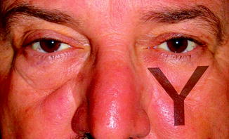
The contour of the midface changes over time. In youth, the midface is full, uniform, and plump. The malar prominence is triangular in shape, with the apex at the junction of the maxilla and zygomatic bones. With increasing age, the midface deflates, leading to the double convexity of the cheek. The nasojugular groove (also known as the tear trough), malar folds, and nasolabial fold become prominent. With time, a characteristic Y-shaped groove develops, created by the palpebromalar groove superolaterally, the nasojugular groove medially, and the midcheek furrow inferolaterally (Fig. ].
Fig. 1.1
The stereotypical Y-shaped groove that develops from the palpebromalar groove superolaterally, the nasojugular groove medially, and the nasojugular groove inferolaterally (Fig. 12 from the chapter Surgical Anatomy of the Forehead, Eyelids, and Midface for the Aesthetic Surgeon by Kevin S. Tan, Sang-Rog Oh, Ayelet Priel, Bobby S. Korn, and Don O. Kikkawa, in Master Techniques in Blepharoplasty and Periorbital Rejuvenation , edited by Dr. Guy Massry, Dr. Azizzdeh, and Dr. Murphy. Springer not yet published)
Osteology
While most aesthetic surgery focuses on the soft tissues of the midface, the underlying bony structures profoundly influence the overall appearance of this area. The significant relationship between skeletal abnormality and facial appearance is evident in the facial deformity in those with maxillary hypoplasia.
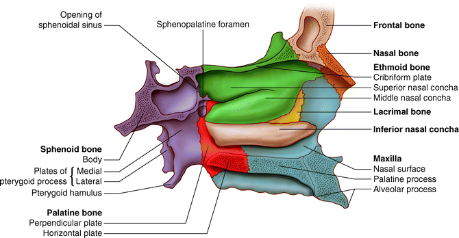
The midface is composed of 14 bones (Fig. ). Six are paired the nasal bones, maxillae, palatine bones, lacrimal bones, zygoma, and inferior nasal conchae. The vomer and mandible compose the remaining bones in the midface. Geometrically, the midfacial bones create a V shape when viewed coronally, as the orbits and zygomatic arches extend beyond the lateral edge of the joined maxillary bones. The maxillary bone sits below the orbits to give the profile of the midface a vertical appearance, with the nasal bones protruding anteriorly.
Fig. 1.2
Coronal view of the 14 bones composing the midface
The paired maxillary bones compose the largest area of the midface and serve as a bridge between the orbits and the mouth. These two bones have the most influence on the overall appearance of the midface. The zygomatic process of the maxilla and the maxillary process of the zygoma meet to form a triangular shape referred to as the malar prominence. The superior portion of each maxilla forms the majority of the orbital floor and contributes to the medial orbital wall as well. The superior two-thirds of the medial edge of each maxilla forms the lateral edge of the nasal aperture. Inferomedially, the two maxillae fuse in the midline to create a platform to house the 16 maxillary teeth. The superior edge of this fusion creates the nasal crest.
Two nasal bones articulate in the midline to create the cephalic portion of the nose. Superiorly, the nasal bones are bordered by the frontal bone. Directly posterior to this, the bones articulate with the ethmoid bone. Posterior and laterally, the frontal process of the maxillary bones join the nasal bones. The medial articulation of the nasal bones, along with the cartilaginous extension beyond the inferior edge of the nasal bones creates the nasal bridge. The anterior nasal aperture is created superiorly by the nasal bones and laterally and inferiorly by the maxillary bones, which meet in the midline of the floor of the nasal cavity to create the nasal crest (Fig. ). Posteriorly, the nasal cavity ends at the posterior nasal aperture, which is created by the paired palatine bone meeting in the facial midline. The nasal cavity is divided in the midline by the nasal septum, which is created by the vomer bone inferiorly and posteriorly. The vomer extends from the perpendicular plate of the ethmoid bone to the nasal spine of the maxillary bone. Superiorly and anteriorly, the nasal aperture is created by the perpendicular plate of the ethmoid bone as it articulates with the nasal bones. The lateral wall of the nasal cavity has contributions from the maxilla, lacrimal, ethmoid, palatine bones, the medial pterygoid plate of the sphenoid bone, and the inferior nasal concha.
Fig. 1.3
Saggittal view of the lateral nasal wall with septum removed
Finally, the mandible articulates with the maxilla at the temporal mandibular joint on either side of the cranium. It is the only facial bone with the ability to move. The mandible houses the 16 lower teeth.