Lucian Fodor , Yehuda Ullman and Monica Elman Aesthetic Applications of Intense Pulsed Light 10.1007/978-1-84996-456-2_1 Springer-Verlag London Limited 2011
1. Skin Anatomy
Lucian Fodor 1, Yehuda Ullmann 1 and Monica Elman 2
(1)
Department of Plastic Surgery, Rambam Health Care Campus, Haifa, Israel
(2)
Maccabbi Insurance, Tel Aviv, Israel
1.1
1.1.1
1.1.2
1.1.3
1.2
1.3
1.3.1
1.3.2
1.3.3
1.4
1.4.1
1.4.2
1.4.3
1.4.4
1.4.5
1.5
1.5.1
1.6
1.7
1.8
Abstract
The skin is composed of three layers: epidermis, dermis and subcutaneous tissue. The thickness of the layers varies with different anatomical regions. The epidermis is thickest on the palm and soles, and very thin on the eyelids, while the dermis is thickest on the back. Keratinocytes are the main component of the epidermis. Melanocytes are the cells located in the epidermis whose function it is to produce pigment. The ratio is about one in every ten basal keratinocytes. Differences in skin color according to race are explained by the number of melanosomes. Langerhans cells represent 35% of the cells of the stratum spinosum where they are situated between the keratinocytes. The dermis consists of a supporting matrix (ground substances) in which polysaccharides and proteins act to produce proteoglycans. The protein fibers inside the dermis are represented by collagen, elastin and other components, such as fibrillin and microfibril proteins. The blood supply to the skin comes from the deep plexuses located at the fascia and subcutaneous level. With aging, there is a decrease in total collagen content in the skin, an increased amount of type III collagen, decreased number and diameter of elastin fibers, and a lack of interaction between water and surrounding molecules which contribute to the dry and wrinkled aspect.
The skin is composed of three layers: epidermis, dermis and subcutaneous tissue. The epidermis is the outer layer and is formed mainly by keratinocytes whose main function is to synthesize keratin. The dermis is the middle layer and its main component is collagen. This layer lies on lobules of lipocytes. The thickness of the layers varies with different anatomical regions. The epidermis is thickest on the palm and soles, and very thin on the eyelids, while the dermis is thickest on the back.
1.1 Epidermis
The epidermis is the outer part of the skin and is composed of three basic cell types: keratinocytes, melanocytes and Langerhans cells. Merkel cells can be found on the palms and soles and are located directly above the basal membrane.
1.1.1 Keratinocytes
Keratinocytes are the main component of the epidermis. Their function is to produce keratin, a complex filamentous protein that forms the stratum corneum of the epidermis.
The epidermis is composed of several layers, beginning with the innermost as follows: basal layer, malpighian layer, granular layer and horny layers (stratum corneum). The palms and soles have also a clear layer called stratum lucidum (above the granular layer). The horny layer and granular layer are the thickest on the palms and soles and are almost absent on the flexor aspect of the forearms. Cycling stem cells, located at the basal layer, provide a pool for epidermal regeneration. As the basal cells divide, they flatten and move upward (Wolff and Wolff-Schreiner ). The process of desquamation implies degradation of the lamellated lipid from the intercellular space and loss of desmosomal interconnections. The keratinocytes play an important role in the immune function of the skin.
1.1.2 Melanocytes
Melanocytes are the cells located in the epidermis whose function it is to produce pigment. The ratio is about one in every ten basal keratinocytes. The face and genitalia have a greater amount of these cells. The melanocyte cell is a dendritic type, extending for long distances within the epidermis and in close contact with the keratinocytes. Together they form the epidermal melanin unit. Melanin is synthetized by melanocytes in the basal layer of the epidermis and transferred to surrounding keratinocytes in melanosomes. Differences in skin color according to race is explained by the number of melanosomes. People with fair skin have fewer melanosomes which are smaller and packaged within membrane complexes. People with darker skin have more melanosomes which are larger and not packed. Sun exposure (Fig ).
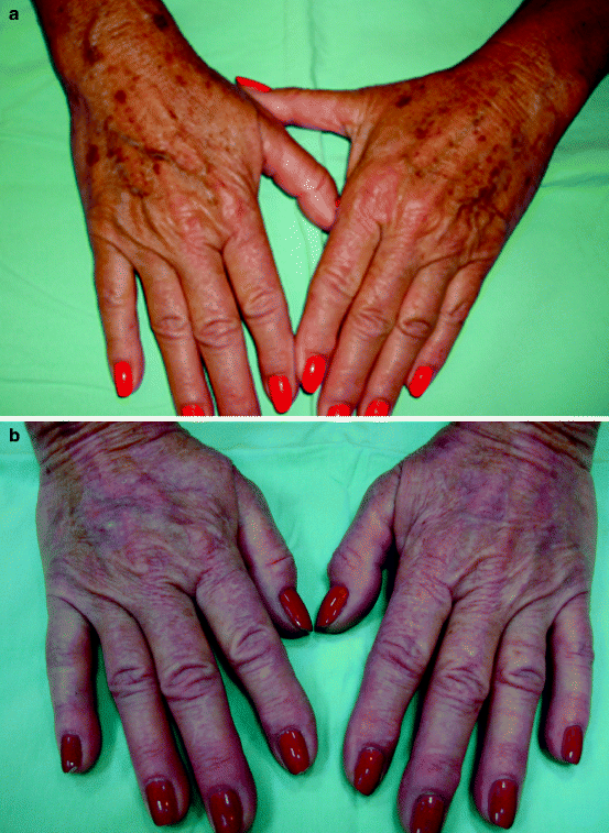
Fig. 1.1
( a ) Significant photoaging and numerous lentigines prior to treatment. ( b ) Eight weeks after a single IPL treatment
1.1.3 Langerhans Cells
Langerhans cells represent 35% of the cells of the stratum spinosum where they are situated between the keratinocytes. They are responsible for the immunological response of the skin.
1.2 Dermoepidermal Junction
The dermoepidermal junction represents the junction between the epidermis and the dermis. It is located at the basement membrane zone and resembles a semi-permeable filter which allows cells and fluids to travel between epidermis and dermis (Briggaman and Wheeler ). It also serves as a structural support for the epidermis.
1.3 Epidermal Appendages
The eccrine and appocrine glands, ducts and pilosebaceous units constitute the skin adnexa. All have a role in epidermis regeneration (reepithelization). When an injury occurs, the keratinocytes from the adnexa migrate to the skin surface (Kollar ).
1.3.1 Eccrine Sweat Glands
These glands have three main components:
The intraepidermal spinal ducts which open directly onto the skin surface
The straight dermal portion of the duct composed of cuboidal epithelial cells
The secretory zone located in the superficial panniculus. In the back region, this zone is situated in the deep dermis
The role of these glands is also to produce sweat which is similar in composition to plasma with regard to the electrolytes. They are important in thermoregulatory function and are present in great amounts in the palms, soles and axillae. Some eccrine glands from the axillae have widely dilated secretory coils in patients with hyperhidrosis.
1.3.2 Appocrine Glands
Appocrine glands develop on the infundibular upper portion of the hair follicle. They are intimally related to the pilar units. The coiled secretory gland is present at the junction of the dermis and subcutaneous fat. Appocrine secretion is odorless and episodic. The appocrine units of the human body are generally confined to the axillae, areolae, genital region, ear canal and eyelids. The glands start to function after puberty.
1.3.3 Hair Follicles
Hair follicles develop in rows of three. Primary follicles are surrounded by the appearance of two secondary follicles. The amount of pilosebaceous units decreases throughout life mainly because of poor formation of secondary follicles. The hair follicle has three main components:
The lower part beginning at the base of the follicle and extending to the insertion of the arrector pili muscle
The middle portion, also called the isthmus, from the arrector pili to the entrance of the sebaceous duct