
This edition first published 2016 2016 John Wiley & Sons, Inc.
Editorial offices: 1606 Golden Aspen Drive, Suites 103 and 104, Ames, Iowa 50010, USA
The Atrium, Southern Gate, Chichester, West Sussex, PO19 8SQ, UK
9600 Garsington Road, Oxford, OX4 2DQ, UK
For details of our global editorial offices, for customer services and for information about how to apply for permission to reuse the copyright material in this book please see our website at www.wiley.com/wiley-blackwell.
Authorization to photocopy items for internal or personal use, or the internal or personal use of specific clients, is granted by Blackwell Publishing, provided that the base fee is paid directly to the Copyright Clearance Center, 222 Rosewood Drive, Danvers, MA 01923. For those organizations that have been granted a photocopy license by CCC, a separate system of payments has been arranged. The fee codes for users of the Transactional Reporting Service are ISBN-13 9781119226260 2016.
Designations used by companies to distinguish their products are often claimed as trademarks. All brand names and product names used in this book are trade names, service marks, trademarks or registered trademarks of their respective owners. The publisher is not associated with any product or vendor mentioned in this book.
The contents of this work are intended to further general scientific research, understanding, and discussion only and are not intended and should not be relied upon as recommending or promoting a specific method, diagnosis, or treatment by health science practitioners for any particular patient. The publisher and the author make no representations or warranties with respect to the accuracy or completeness of the contents of this work and specifically disclaim all warranties, including without limitation any implied warranties of fitness for a particular purpose. In view of ongoing research, equipment modifications, changes in governmental regulations, and the constant flow of information relating to the use of medicines, equipment, and devices, the reader is urged to review and evaluate the information provided in the package insert or instructions for each medicine, equipment, or device for, among other things, any changes in the instructions or indication of usage and for added warnings and precautions. Readers should consult with a specialist where appropriate. The fact that an organization or Website is referred to in this work as a citation and/or a potential source of further information does not mean that the author or the publisher endorses the information the organization or Website may provide or recommendations it may make. Further, readers should be aware that Internet Websites listed in this work may have changed or disappeared between when this work was written and when it is read. No warranty may be created or extended by any promotional statements for this work. Neither the publisher nor the author shall be liable for any damages arising herefrom.
Library of Congress Cataloging-in-Publication Data applied for.
ISBN: 9781119226260
A catalogue record for this book is available from the British Library.
Wiley also publishes its books in a variety of electronic formats. Some content that appears in print may not be available in electronic books.
Preface: Why maximizing your biopsy results is important
A biopsy is the collection of tissue to be analyzed by the pathologist. It is often the gold standard of diagnosis. Ideally, from the pathologist's perspective, all lesions would be biopsied so that treatment could be based on a confirmed diagnosis. However, as with most decisions, the decision to perform a biopsy should be based on a cost-benefit analysis. The costs include time, biopsy procedure fee, histology fee, pet discomfort, possible infection or spread of disease, and owner inconvenience. The potential benefits are an accurate diagnosis and prognostic information to guide future treatment, decrease patient suffering, and avoid the cost of treating an assumed incorrect condition. There are many steps in the biopsy process that can be optimized to ensure minimizing the cost and maximizing the benefit. This manual is designed to help clinicians submit cost-effective biopsies by providing suggestions for each step of the biopsy submission, separate organ specific biopsy considerations, a step-by-step biopsy submission checklist, and a list of general biopsy dos and don'ts. It is hoped that this manual provides enough detail to be helpful, but not so much that the useful information is obscured, and that it will be consulted often prior to biopsy collection.
Acknowledgments
I gratefully acknowledge Drs. Thomas Lipscomb and Frances Moore for helpful comments on the manuscript, Dr. Anne Kincaid for helping to collect case material, Ms. Ingrid Style for her advice on the drawings, Mr. Lance Schuette and Ms. Pam Schmidt for scanning slides, Marshfield Laboratory histology technicians for some of the gross photos, Dr. Michelle Fleetwood for help with the literature review, and Julie, Mary, and Charles Lipscomb for their encouragement.
Chapter 1
Steps of a Successful Biopsy Submission
1 Collection
Each step of the biopsy submission process is important and contributes to the accuracy of the diagnosis, but the collection step is critical. While submission form information can be added to or revised at a later date and the correlation between the histologic and clinical assessments can be reassessed as new information is provided, the quality of the biopsy specimen is irrevocably determined by the collection method. One cannot make up for improper collection or fixation with a longer clinical history.
i Site
When presented with a single, small lesion, the site selection is straightforward (). Be sure to biopsy any intact vesicles or pustules. For lesions that are too large to excise, multiple biopsies, including all grossly different appearing areas, are recommended in an attempt to submit fully representative specimens.
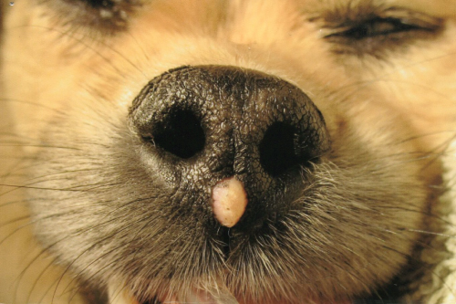
Solitary mass, viral papilloma, on the nose of a terrier. A single excisional biopsy was diagnostic and curative. (Reproduced by permission of Bass Lake Pet Hospital, New Hope, MN 55428).
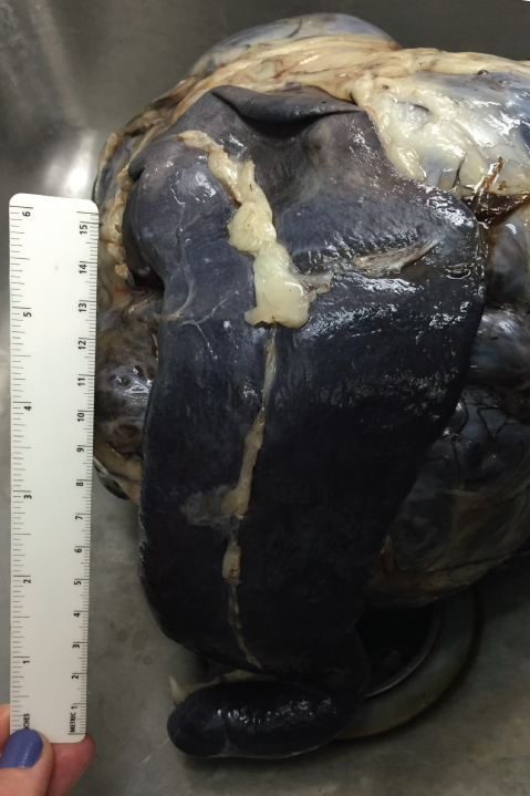
Spleen (in the foreground) with a large mass attached to the far side that requires sampling of all grossly different appearing areas to fully evaluate the lesion.
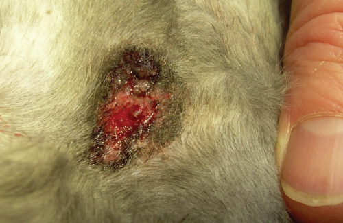
Ulcerated skin lesion (feline squamous cell carcinoma in situ [Bowen's-like disease] in this case). Excisional biopsy or sampling the periphery of the lesion is more likely to be diagnostic than a biopsy of the central ulcerated area. (Reproduced by permission of Whitewater Veterinary Hospital, Whitewater, WI 53190).
ii Size
While the type of biopsy specimen collected (such as needle, punch, incisional or excisional biopsies (). In other words, the larger the biopsy sample, the more likely it is to be fully representative and diagnostic. When clinically reasonable, excisional biopsies are recommended. Not only are excisional biopsies fully representative of the lesion, but they can also be curative.
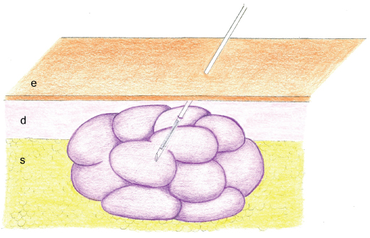 Next page
