1. Barrier Skin
The skin is a major interface between the body and the environment. In most cases the skin serves as a perfect barrier; however it is dependent on several factors such as the chemical properties of a compound to which the skin is exposed topically, but also its concentration, the contact duration, frequency of exposure, and the exposed surface area influence the amount of penetration which may lead to local reactions including irritation, sensitization, and inflammation but also to penetration and entering the systemic circulation which may result in systemic effects [].
There are several lines of defenses by the barrier organ skin:
The chemo-physical barrier of the stratum corneum
The immunocompetent cells of the epidermis and corium
The armamentarium of xenobiotica-metabolizing enzymes in epidermal cells such as the keratinocytes as well as antigen-presenting cells, e.g., the Langerhans cells
The melanocyte-keratinocyte unit and its role in pigmentation and protection against UV radiation
The main chemo-physical barrier of the skin is located in the outermost layer of the skin, the stratum corneum, which consists out of corneocytes surrounded by lipid regions. The main protein are the keratins; however they need several other proteins for their formation in a way that they can function as a barrier. One of these proteins is filaggrin. Filaggrin is a protein which therefore is essential for a normal barrier formation in stratum corneum, and a diminished expression of this protein is associated with the precipitation of atopic dermatitis and ichthyosis vulgaris if the mutation of filaggrin is homozygous and no active filaggrin results [].
Table 1.1
Xenobiotica with noticeable local or systemic toxicity []
Herbicides |
Paraquat |
Monochloroacetic acid |
Pesticides/insecticides (organophosphates) a |
Methyl bromide |
Malathion |
Strychnine |
Amitraz |
Pyrethroids |
Chlorpyrifos |
Diazinon |
N,N-Diethyl-m-toluamide |
Cutaneous medications |
Topical -blockers |
Topical 2-agonists |
Dorzolamide/brinzolamide |
Topical prostaglandin analogs |
Minoxidil |
Nitroglycerin |
Resorcinol |
Tretinoin |
Adapalene |
Salicylic acid |
Sodium sulfacetamide |
Nicotine |
Glucocorticoides |
Occupational contact |
Trichloroethylene |
Lindane |
Chromium |
Engine oil |
Hydrofluoric acid |
Isocyanates |
Plasticizers (most commonly phthalate esters)b |
Cosmeceuticals and Diversa |
Mercury |
Phenol |
Arsenic |
aAlone in the USA, >40 organophosphate pesticides are registered, and EPA estimates that in 2007 about 33 million pounds were applied []
bPhthalate esters are ubiquitous and are used to manufacture building materials, household furnishings, clothing, cosmetics, pharmaceuticals, nutritional supplements, medical devices, dentures, childrens toys, glow stick, modeling clay, food packaging, automobiles, lubricants, waxes, cleaning materials, and insecticides []
The stratum corneum is not only a barrier for most xenobiotica but also for physical agents such as UV light. Under its influence the differentiation of keratinocytes is altered in a way that the stratum corneum is enlarged which improves its capability to absorb UV light.
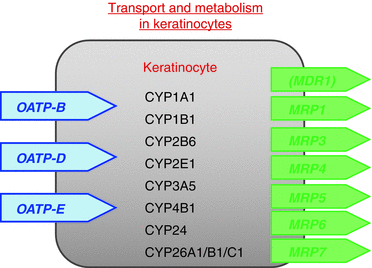
The stratum corneum is a perfectly specialized chemo-physical barrier and an umbrella over the epidermis. But also the epidermis functions as a barrier because it works as an immunologic first line of defense and xenobiotica-metabolizing organ. In addition it protects the skin against the damages by UV radiation. The epidermis possesses several highly specialized cells which are connected by several interacting signaling processes with one another. The main compartment are the keratinocytes, which do not only serve as a source for the corneocytes in the stratum corneum compartment, but they contribute to inflammatory processes, e.g., by the production and release of multiple cytokines which modulate inflammatory processes. Keratinocytes play a major role in the synthesis of vitamin D and are able to metabolize xenobiotica by enzymes such as the cytochrome P450 isoenzymes which are inducible by xenobiotica (Fig. ).
Fig. 1.1
Cytochrome P450 (CYP) and influx (OATP) as well as efflux proteins (MDR/MRP) which have been characterized in human keratinocytes []
Table 1.2
Xenobiotica metabolizing enzymes in the skin
Xenobiotica-metabolizing enzymes in the skin |
|---|
Cytochrome P450 |
Flavin-dependent monooxygenases (FMO) |
Cyclooxygenases (COX1/COX2) |
Alcohol dehydrogenase (ADH) |
Aldehydedehydrogenase (ALDH) |
NAD(P)H:quinone reductase (NQR) |
Epoxide hydrolase (EH) |
Esterase/amidase |
Glutathione S-transferase (GST) |
UDP-glucuronosyltransferase (UGT) |
Sulfotransferase (SULT) |
N-Acetyltransferase (NAT) |
Melanocytes are dendritic cells, which produce melanin, which is transported to connected keratinocytes upon UV radiation, and this melanocyte together with about 3040 keratinocytes forms a melanocyte-keratinocyte unite, which plays a major role in the UV protection of the skin. The induction of the vitamin D synthesis in keratinocytes by UVB light also augments the expression of filaggrin which improves the barrier formation by the stratum corneum [].
Further on the skin possesses immunocompetent cells including antigen-presenting dendritic cells and T-lymphocytes which play an important role as an immunologic first line of defense but may also lead to sensitization and inflammatory responses to environmental hazards [].
The pathophysiology of two inflammatory skin diseases which belong to the most often precipitated disorders of the skineczema and psoriasisis closely associated with dysfunctions of the skin barrier. The role of filaggrin in the pathophysiology of atopic dermatitis has been mentioned already. The precipitation of both pathological symptomseczema and psoriasisis dependent on Th1-mediated immunological processes. In the case of eczema formation, one differentiates between atopic dermatitis, which are most often triggered by Th2-dependent allergic reactions, and after a switch of the infiltrating T-lymphocytes to Tc1-type lymphocytes, eczema is precipitated as in the case of allergic contact dermatitis which is triggered and precipitated by Tc1-type lymphocytes []. Atopic dermatitis is quite often triggered by protein antigens originating from, e.g., house dust mite, food allergens, and others, whereas allergic contact dermatitis is triggered by small molecular weight compounds.

