Elastographic images rely on two different physical approaches: the assessment of the strain produced in the tissue by external compression (strain imaging) or the evaluation of the shear waves (SW) speed propagation in the medium (shear wave imaging). Both methods aim to evaluate qualitatively (display contrast) and quantitatively (measure) the Youngs modulus (YM). Despite the YM does not take into account the system size and the amount of material, it is considered the physical parameter corresponding to stiffness [].
In biological tissues two different mechanical properties act against any type of deformation (shear): elasticity and viscosity [].
1.1.1 Strain Compression Imaging
In strain imaging compression (stress) could theoretically be applied in three different ways: on the whole surface, peripherally on a surface, or homogeneously on a volume. Each of them is described by his own modulus: YM, shear modulus, and bulk modulus, respectively. In order to simplify the comprehension, we will focus our discussion on the two-dimensional model avoiding a detailed explanation of the bulk modulus.
As previously said a simple shearing force can be applied to a single portion of an object causing a deformation without volume modification (shear stress, SS). Contrary a deformation could be the result of a compressive force on the whole surface of the body (YM) [];
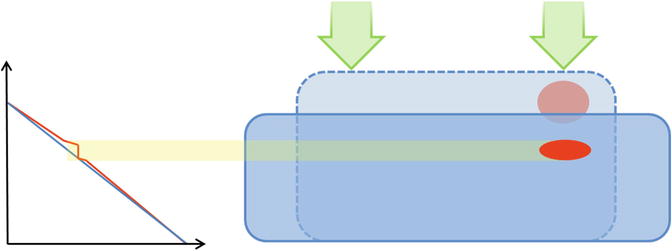
YM evaluation model: if an imaginary rectangular shape of a homogeneous material is rigidly bound along one face and further compressed by a parallel plate on the other, its form will be modified and expanded along the axis perpendicular to the one of the compression force, but its volume will be maintained [] which depends on the different characteristics of the other materials.
Fig. 1.1
Exemplification of the pure stress ( arrows ) applied on the surface of a rectangular shaped homogeneous medium, which deforms it maintaining its volume. The blue line on the diagram represents the graphic correlation, whereas the red line depicts the modification related to the presence of an inhomogeneity within the same material
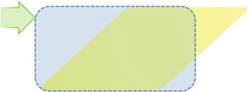
SS evaluation model: applying the same force but on a different direction (perpendicular to the former and on one edge) to the same imaginary rectangular-shaped model, the block will again modify its form without any volume variation. Once again images will be obtained at different times, previous and during material modification, and the presence of inclusions will perturb the graphical representation in the same way as explained before.
Fig. 1.2
Exemplification of a shear stress ( arrow ) applied on an edge of the rectangular shaped homogeneous medium deforms it maintaining its volume
In the purely elastic module described above, stress and strain are linked through the Hooks law. The equation which explains this relation is

, where is the stress applied, E the YM, and = L / L the longitudinal strain. The deformations occurring in human tissues are far more complex than what explained here, but these two ideal cases approximate the basis of the two imaging modalities: SS exemplifies ARFI imaging, whereas YM describes those produced by transducer compression.
1.1.2 Shear Wave Imaging
In SW imaging, a time-varying force is applied to the tissue. This force can be a limited transient mechanical force or a fixed frequency oscillatory one []. Therefore, SW imaging is based on the propagation of SWs which propagate in the tissues perpendicularly to the ordinal pulse echo longitudinal waves.
SWs are slow transverse waves which attenuate rapidly compared to the diagnostic longitudinal waves and disappear in the MHz ultrasound band, with a propagation frequency below 1 KHz in vivo. Their velocity is 1000 times slower (i.e., c s = 110 m/s) compared to longitudinal waves ( c L = 1540 m/s) [].
When the free face of the rectangular shape described above is displaced repetitively, the behavior of the medium obeys to classic wave equation [].
All these physical theories and models allowed the development of multiple methods, which have been integrated in clinical practice:
Strain elastography (SE)
Transient elastography (TE)
ARFI
SW speed measurement and imaging
Strain elastography
Tissue displacement is produced by tissue compression with the probe. Since static compression is subjectively achieved, only strain () is displayed; as a consequence, this type of imaging is only qualitative. Alternatively, pseudo-quantitative methods such as the strain ratio [].
Transient elastography
A controlled vibrating external piston (which acts as a punch) mounted on a probe with a fixed focal spot is used to either generate or evaluate the SW which has been produced. This type of SW quantification is used essentially by FibroScanTM to evaluate tissue stiffness.
ARFI
A focused acoustic radiation force pushing pulse is used to deform tissues within a defined region. The probe works as a generator of the push and to monitor tissue displacement sending an imaging pulse prior and after the push impulse. The evaluation of multiple beam lines allows the creation of an image, which displays the intrinsic differences of the tissue.













 , where is the stress applied, E the YM, and = L / L the longitudinal strain. The deformations occurring in human tissues are far more complex than what explained here, but these two ideal cases approximate the basis of the two imaging modalities: SS exemplifies ARFI imaging, whereas YM describes those produced by transducer compression.