• Complain

Thuy L. Phung - Pediatric Dermatopathology

Here you can read online Thuy L. Phung - Pediatric Dermatopathology full text of the book (entire story) in english for free. Download pdf and epub, get meaning, cover and reviews about this ebook. year: 2017, publisher: Springer International Publishing, Cham, genre: Home and family. Description of the work, (preface) as well as reviews are available. Best literature library LitArk.com created for fans of good reading and offers a wide selection of genres:

Romance novel Science fiction Adventure Detective Science History Home and family Prose Art Politics Computer Non-fiction Religion Business Children Humor

Choose a favorite category and find really read worthwhile books. Enjoy immersion in the world of imagination, feel the emotions of the characters or learn something new for yourself, make an fascinating discovery.

Thuy L. Phung Pediatric Dermatopathology

Pediatric Dermatopathology: summary, description and annotation

We offer to read an annotation, description, summary or preface (depends on what the author of the book "Pediatric Dermatopathology" wrote himself). If you haven't found the necessary information about the book — write in the comments, we will try to find it.

Thuy L. Phung: author's other books


Who wrote Pediatric Dermatopathology? Find out the surname, the name of the author of the book and a list of all author's works by series.

Pediatric Dermatopathology — read online for free the complete book (whole text) full work

Below is the text of the book, divided by pages. System saving the place of the last page read, allows you to conveniently read the book "Pediatric Dermatopathology" online for free, without having to search again every time where you left off. Put a bookmark, and you can go to the page where you finished reading at any time.

Light

Font size:

Reset

Interval:

Bookmark:

Make
Springer International Publishing Switzerland 2017
Thuy L. Phung , Teresa S. Wright , Crystal Y. Pourciau and Bruce R. Smoller Pediatric Dermatopathology 10.1007/978-3-319-44824-4_1
1. Spongiotic Dermatitis
Thuy L. Phung 1, Teresa S. Wright 2, Crystal Y. Pourciau 3 and Bruce R. Smoller 4
(1)
Department of Pathology & Immunology, Baylor College of Medicine and Texas Childrens Hospital, Houston, Texas, USA
(2)
Departments of Dermatology and Pediatrics, University of Tennessee Health Science Center, Memphis, TN, USA
(3)
Departments of Dermatology and Pediatrics, Baylor College of Medicine and Texas Childrens Hospital, Houston, Texas, USA
(4)
Department of Pathology and Laboratory Medicine, University of Rochester School of Medicine and Dentistry, Rochester, NY, USA
1.1 Atopic Dermatitis
1.1.1 Clinical Features
Atopic dermatitis is a multifactorial, primarily type 2 T-helper cell immune-mediated, inflammatory disorder characterized by chronicity, xerosis, and pruritus. Sixty percent of cases present within the first year of life, with 90 % of patients demonstrating clinical disease by 5 years old []. There is a strong genetic predisposition with 70 % of patients having a positive family history of atopy.
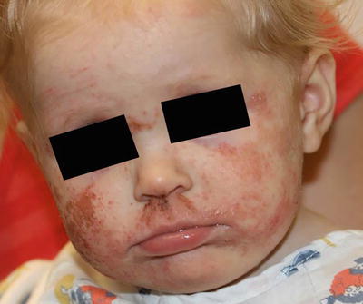
Clinical examination shows erythematous, xerotic, scaled patches and plaques with or without weeping crust and overlying excoriations. Lichenification and exaggerated skin markings highlight disease chronicity. In infants, eczematous lesions are found on the face and extensor surfaces with sparing of the diaper area, owing to pronounced humidity at that site. In contrast, older children and adults usually have skin findings localized to the flexural skin of the neck, antecubital fossae, wrists, popliteal fossae, dorsum of the feet and ankles (Fig. ).
Fig 11 Atopic dermatitis presents as erythematous scaly plaques over the - photo 1
Fig. 1.1
Atopic dermatitis presents as erythematous, scaly plaques over the face of a toddler
Atopic dermatitis is described as the first step of the atopic march with 4060 % of patients later developing allergic rhinoconjunctivitis and/or asthma [].
1.1.2 Histology
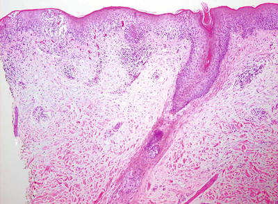
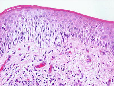
Histologic changes in acute atopic dermatitis are quite subtle. Scant wispy parakeratosis overlies an epidermis that demonstrates slight spongiosis and small collections of Langerhans cells [].
Fig 12 Atopic dermatitis presents with a mild spongiotic dermatitis and a - photo 2
Fig. 1.2
Atopic dermatitis presents with a mild spongiotic dermatitis and a mild lymphocytic infiltrate in the superficial dermis
Fig 13 A spongiotic dermatitis with scant exocytosis of lymphocytes is seen - photo 3
Fig. 1.3
A spongiotic dermatitis with scant exocytosis of lymphocytes is seen in atopic dermatitis
In the chronic phase, changes are nonspecific and are those of a chronic spongiotic dermatitis, often with superimposed lichen simplex chronicus-like changes. Hyperkeratosis is present diffusely overlying an acanthotic epidermis with elongated rete ridges and hypergranulosis. Spongiosis is only minimally present and there is a sparse perivascular lymphocytic infiltrate. Eosinophils are minimally increased in number. A specific diagnosis of chronic atopic dermatitis is not possible based solely upon histologic changes .
1.1.3 Pathogenesis
Genetics studies have revealed a strong association between atopic dermatitis and the epidermal barrier protein filaggrin , particularly in Northern Europeans []. These variants also show significant association with asthma in the setting of atopic dermatitis.
The filaggrin gene is located within the epidermal differentiation complex, which is a large cluster of genes (including filaggrin, loricrin, and involucrin) that are important in keratinocyte terminal differentiation [].
The antimicrobial skin barrier is compromised in atopic dermatitis, leading to secondary bacterial infection, such as Staphylococcus aureus colonization that can exacerbate inflammation in the disease process [].
T lymphocytes play important roles in the pathogenesis of atopic dermatitis. The important T lymphocyte subsets in this disorder are type 2 T-helper cells (Th2) , type 17 T-helper cells (Th17) , and type 22 T-helper cells (Th22) [].
Vascular changes and angiogenesis are other key features in atopic dermatitis. High levels of vascular endothelial growth factor-A (VEGF-A) are present in the skin of atopic dermatitis patients and appear to correlate with disease activity []. Activated macrophages can induce VEGF expression and angiogenesis during inflammation and contribute to the disease process.
Atopic dermatitis can occur as part of a syndrome, such as Netherton syndrome which is characterized by ichthyosis, chronic dermatitis, asthma, and allergic rhinitis [].
1.2 Dyshidrotic Eczema
1.2.1 Clinical Features
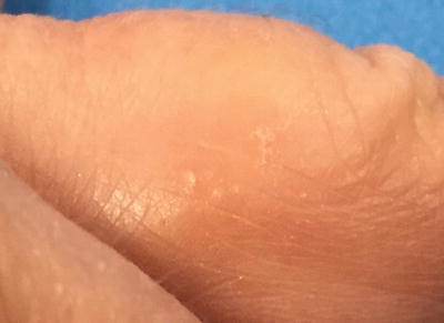
Dyshidrotic eczema, also known as pompholyx, is a chronic relapsing, vesiculobullous disorder of the palms and soles (Fig. ].
Fig 14 Dyshidrotic eczema presents as scaly papules and deep-seated vesicles - photo 4
Fig. 1.4
Dyshidrotic eczema presents as scaly papules and deep-seated vesicles on the lateral finger
On physical exam, there are tense, deep-seated, tapioca-like vesicles as well as larger bullae on the palms, soles, and lateral fingers. Erythema and scaling may or may not be present. Involvement is usually bilateral. Patients may complain of pruritus or a burning sensation, and hyperhidrosis is a frequent feature, typically with increased occurrence during warmer seasons and climates []. Management hinges on avoidance of identifiable triggers and treatment of relapses of eczematous flares.
1.2.2 Histology
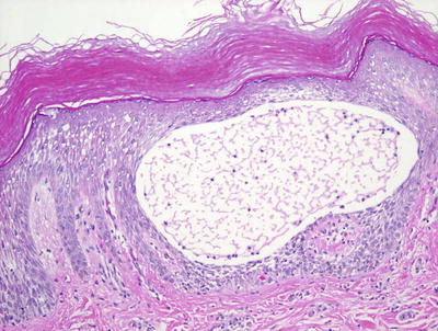
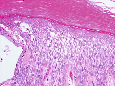
Histologic features of dyshidrotic eczema are nonspecific. The only distinguishing feature from other spongiotic processes is the acral location for this eruption (Fig. ). Within the dermis, there is an infiltrate of lymphocytes and eosinophils may be present. Late lesions may demonstrate only minimal spongiosis and are indistinguishable from lichen simplex chronicus. In most cases, a PAS stain is recommended to exclude acral fungal infection, as the histologic changes may be identical .
Fig 15 Acral skin demonstrating spongiotic dermatitis with microvesiculation - photo 5
Fig. 1.5
Acral skin demonstrating spongiotic dermatitis with microvesiculation is present in dyshidrotic eczema
Fig 16 Dyshidrotic eczema reveals a spongiotic dermatitis on acral skin - photo 6
Fig. 1.6
Dyshidrotic eczema reveals a spongiotic dermatitis on acral skin
1.2.3 Pathogenesis
The pathogenesis of dyshidrotic eczema is not known. A number of factors, such as atopy, contact allergy, dermatophyte infection and drug eruption, can influence the development of dyshidrotic eczema in predisposed individuals. Studies of a large Chinese family with a rare autosomal dominant form of dyshidrotic eczema have identified a locus on chromosome 18q22.118q22.3 that may contain the responsible gene(s) []. There are 17 genes within this locus, some of which are calcium-dependent adhesion molecules, such as cadherins, which are important in maintaining the structural integrity of epithelial tissues including the skin .
Next page
Light

Font size:

Reset

Interval:

Bookmark:

Make

Similar books «Pediatric Dermatopathology»

Look at similar books to Pediatric Dermatopathology. We have selected literature similar in name and meaning in the hope of providing readers with more options to find new, interesting, not yet read works.


Reviews about «Pediatric Dermatopathology»

Discussion, reviews of the book Pediatric Dermatopathology and just readers' own opinions. Leave your comments, write what you think about the work, its meaning or the main characters. Specify what exactly you liked and what you didn't like, and why you think so.