Embryology of the Skin
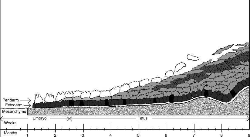
The youngest human embryos in which cutaneous development was observed were about 45 weeks old. At that point, the embryos external surface is covered by the ectoderm, a monolayer of cells stretching over a mesenchymal mantle. It is partially overlaid by the periderm, a second layer of bulky elements endowed with microvilli. This situation and ensuing events are presented in Fig..
Fig. 1.1
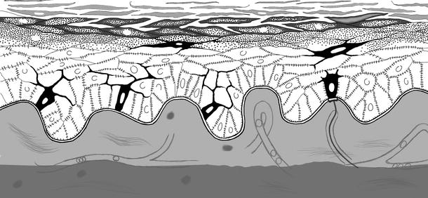
Diagrammatic representation of the development of human skin. The germinal layer is depicted in heavy gray , and the melanocytes interspersed in it are shown in black . The granular layer is represented as a dark gray dotted band while desmosomes are pictured as faint interstitial lines . The basement membrane appears as a heavy double line . For the sake of simplicity, Langerhans and Merkel cells, as well as nuclear contours, are omitted from the drawing. The time of gestation in weeks and months is indicated along the horizontal axis of the figure
The ectodermal layer develops into the stratum germinativum, a permanent element which evolves into the germinal layer of postnatal life, whereas the periderm, a decidual layer, flattens and is ultimately shed into the amniotic cavity. By the tenth week of gestation, when the embryo turns into a fetus, an intermediate layer of cells develops from the stratum germinativum beneath the periderm. This is known as the stratum intermedium. This layer, which corresponds to the stratum spinosum of postuterine life, proliferates and, by week 23, undergoes keratinization. This event is evidenced by the appearance of keratohyalin granules; the development of the granular layer is coincident with the loss of the periderm into the amniotic cavity.
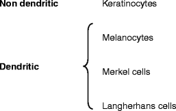
By week 67, a basement membrane appears and soon attains considerable thickness. Melanocytes reach the epidermis by the 12th week of gestation, remaining from that point on in the stratum germinativum. The origin of the Merkel cells is not well established, but the presence of desmosomes among them and the surrounding keratinocytes suggests that they may differentiate from the latter. Langerhans cells, the fourth cellular component of the epidermis, are generated in the bone marrow, from where they migrate and colonize the epidermis.
Embryology of the Cutaneous Adnexa
Toward the 12th week of gestation, primary epithelial germs develop as condensations on the underface of the stratum germinativum. Some of these, which then give rise to the pilosebaceous/apocrine complex, protrude into the dermis as pilar germs, as part of a process induced by mesenchymal cells beneath. Later, when the pilar papilla of mesenchymal origin intrudes into the basis of the pilar germs, they are transformed into bulbopilar germs. As the bulbopilar germs keep growing downward, two bulges emerge from their exterior: a superior one corresponding to the origin of the sebaceous gland and an inferior one linked to the insertion of the arrector pilorum . In certain areas of the body, the bulbopilar germs exhibit a top third bulge corresponding to the origin of the apocrine gland. The central part of the bulbopilar germ is transformed into the hair shaft and the peripheral part into the inner radicular sheath, later to be surrounded by the outer radicular sheath, of epidermal origin.
The eccrine glands originate from primary epithelial germs, independently from the bulbopilar ones. The ungual complex appears upon the 12th week of gestation as epidermal thickenings at the dorsum of the fingertips, being laterally limited by two folds, and at the base by the proximal fold, from beneath which the nail matrix develops.


