Kasia S. Masterpol - Atlas of Essential Dermatopathology
Here you can read online Kasia S. Masterpol - Atlas of Essential Dermatopathology full text of the book (entire story) in english for free. Download pdf and epub, get meaning, cover and reviews about this ebook. year: 2012, publisher: Springer Nature, genre: Romance novel. Description of the work, (preface) as well as reviews are available. Best literature library LitArk.com created for fans of good reading and offers a wide selection of genres:
Romance novel
Science fiction
Adventure
Detective
Science
History
Home and family
Prose
Art
Politics
Computer
Non-fiction
Religion
Business
Children
Humor
Choose a favorite category and find really read worthwhile books. Enjoy immersion in the world of imagination, feel the emotions of the characters or learn something new for yourself, make an fascinating discovery.
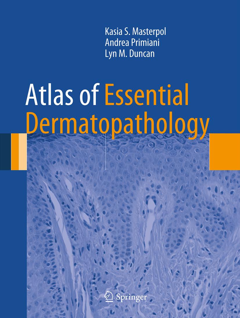
- Book:Atlas of Essential Dermatopathology
- Author:
- Publisher:Springer Nature
- Genre:
- Year:2012
- Rating:5 / 5
- Favourites:Add to favourites
- Your mark:
Atlas of Essential Dermatopathology: summary, description and annotation
We offer to read an annotation, description, summary or preface (depends on what the author of the book "Atlas of Essential Dermatopathology" wrote himself). If you haven't found the necessary information about the book — write in the comments, we will try to find it.
Kasia S. Masterpol: author's other books
Who wrote Atlas of Essential Dermatopathology? Find out the surname, the name of the author of the book and a list of all author's works by series.


Atlas of Essential Dermatopathology — read online for free the complete book (whole text) full work
Below is the text of the book, divided by pages. System saving the place of the last page read, allows you to conveniently read the book "Atlas of Essential Dermatopathology" online for free, without having to search again every time where you left off. Put a bookmark, and you can go to the page where you finished reading at any time.
Font size:
Interval:
Bookmark:
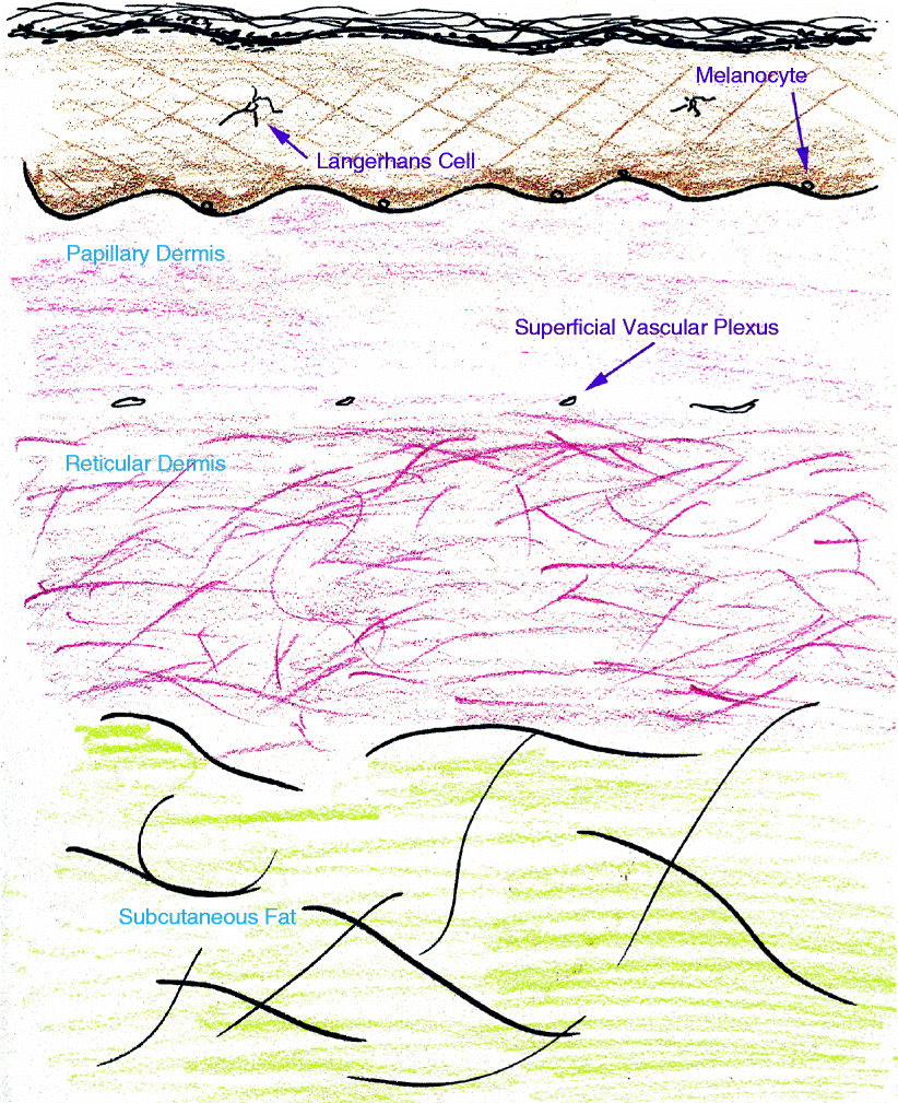
Anatomy
- Stratified squamous epithelium composed of layers of keratinocytes
- From superficial to deep:
- Stratum corneum
- Stratum granulosum
- Stratum spinosum
- Stratum basale
- Intermingled cell types :
- Melanocytes: at dermoepidermal junction
- Transfer melanin keratinocytes
- Langerhans cells: CD1a + and Langerin + dendritic cells in stratum spinosum
- Function in antigen presentation
- Merkel cells: neuroendocrine cells in the stratum basale, associated with nerve endings from the dermis
- Papillary dermis :
- Dermal papillae complement rete ridges of epidermis
- Fine, pale eosinophilic collagen fibers
- Contains free nerve endings and Meissners corpuscles
- Separated from the reticular dermis by the superficial vascular plexus
- Reticular dermis :
- Thick, deeply eosinophilic collagen fibers
- Contains the deep vascular plexus, adnexal structures, nerve trunks, Pacinian corpuscles, glomus bodies
- Separated into lobules by fibrous septae extending from the reticular dermis
- Contains anagen hair bulbs and medium sized arterioles and veins
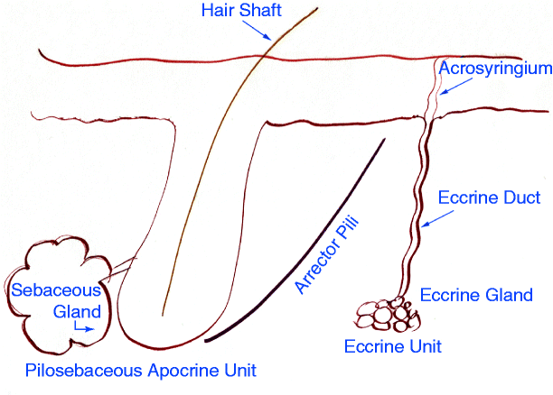
- Eccrine Glands : palms, soles, forehead, axillae
- Coiled, secretory component in deep dermis
- Single layer of cuboidal epithelium, eosinophilic cytoplasm
- Surrounded by myoepithelial cells
- Eccrine Ducts
- Long duct, extends from glandular coil in deep dermis to exit through the epidermis as an acrosyringium
- Two layers of epithelium, no myoepithelial cells
- Hair Follicles
- Types: terminal (diameter 0.06 mm), vellus (diameter 0.03 mm)
- Zones (from superficial to deep):
- Infundibulum = region above entry of sebaceous gland duct
- Isthmus = extends from attachment of arrector pili muscle to entry of sebaceous gland duct
- Hair bulb = dermal papillae and hair matrix
- Phases: Anagen (growth), Catagen (involution), Telogen (resting phase)
- Hair shaft = composed of cuticle, cortex, and medulla
- Arrector pili = smooth muscle innervated by sympathetic nervous system
- Sebaceous Glands
- Acinar pattern, multiple lobules
- Inner layers of cells with vacuolated, lipid-filled cytoplasm
- Outer rim of cuboidal basophilic germinative cells
- Short duct with stratified squamous epithelium, enters into pilosebaceous unit
- Rarely the sebaceous duct exits through the epidermis directly
- Apocrine Units : axillae, anogenital region, areola, eyelid
- Coiled, secretory component in dermis
- Decapitation secretion, snouts
- Single layer of cuboidal to columnar epithelial cells, eosinophilic cytoplasm
- Surrounded by myoepithelial cells
- Short duct opens into infundibulum of associated hair follicle
Font size:
Interval:
Bookmark:
Similar books «Atlas of Essential Dermatopathology»
Look at similar books to Atlas of Essential Dermatopathology. We have selected literature similar in name and meaning in the hope of providing readers with more options to find new, interesting, not yet read works.
Discussion, reviews of the book Atlas of Essential Dermatopathology and just readers' own opinions. Leave your comments, write what you think about the work, its meaning or the main characters. Specify what exactly you liked and what you didn't like, and why you think so.